Full Mouth Rehabilitation in Patient with Dentinogenesis Imperfecta in Primary Dentition: A Case Report
Department of Pedodontics & Preventive Dentistry, Sri Aurobindo College of Dentistry, Indore.(MP)
ABSTRACT
Background - Dentinogenesis imperfecta or hereditary opalescent dentin was first described in the late 19th century. It is a localized mesodermal dysplasia affecting both the primary and permanent dentition.
Case report- A 5-year-old/ male medically compromised patient reported to the department with complaint of difficulty in chewing. On examination deciduous teeth showed generalized brownish discolouration, severe attrition and dehiscence. Full mouth prosthetic rehabilitation was done for the patient it helped him to improve his aesthetics, rebuild his self-confidence and improve the quality to his life.
Conclusion- Full mouth rehabilitation improved aesthetics, thus has helped in developing positive psychological support for the child. Children with DI can be saved from peer harassment resulting in increased self-esteem and confidence.
INTRODUCTION
Dentinogenesis imperfecta or hereditary opalescent dentin, was first described in the late 19th century.1 It is a localized mesodermal dysplasia affecting both the primary and permanent dentition. It is defined as “A hereditary developmental disturbance of the dentin originating during the histodifferentiation stage of tooth development.”2 It was found to be the rarest dental anomaly with a prevalence of 0.09% in the city of Indore, India.3
It may be seen alone or in conjunction with the systemic hereditary disorder of the bone, Osteogenesis Imperfecta. The other two types are autosomal dominant conditions that have been linked to allelic mutations of the DSPP gene encoding dentin phosphoprotein and dentin sialoprotein.4,5
The treatment goals for treating such patients with primary dentition are:
- Maintain Dental Health by preserving vitality, form, and size of the dentition.
- Enhance Esthetic to Prevent psychological issues.
- Provide Functional Dentition which helps in chewing.
- Prevents loss of vertical dimension.
- Maintains the arch length.
- Avoid interfering with the eruption of the remaining permanent teeth.8
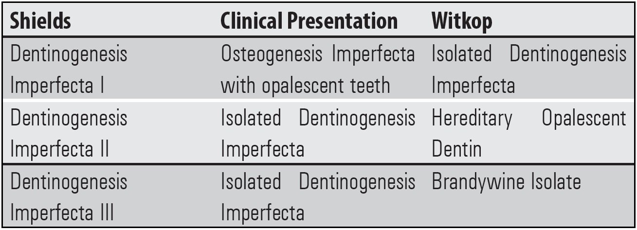
Fig 1
The two most accepted classifications are given by Shield and Witkop.6,7
CASE REPORT
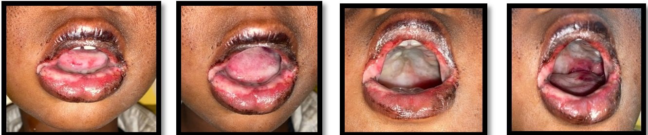
5-year-old/ male patient reported to the department of Paediatric and Preventive Dentistry of Sri Aurobindo College of Dentistry with the presenting complaint of inability to eat food due to difficulty in chewing, brown discoloration of all his teeth since birth and excessive wearing of teeth. Patient gave a medical history of viral hepatitis, nutritional anemia, developmental delay, and speech delay. No history of bone fracture was given. Intra-oral examination showed generalized brownish discoloration with primary dentition, severe attrition with 53,52,61,63,64,72,73,74,75,82,83,84,85;dehiscence with 54,55,64,65,75; Greyish discoloration with 16,26,36,31,41,46; Root Stumps were present with 51,62 ; 31,41 were seen erupting. (FIGURE 1a, 1b) Extraoral examination revealed developing class III malocclusion.

Radiographic examination (Figure 2) revealed short & conical roots with peri-apical radiolucency. The coronal structure had wide pulp chambers and cervical constriction. Generalised severe boneless was noted. The Final diagnosis of Dentinogenesis Imperfecta type 2 was given on the basis of clinical and radiographic assessment. A comprehensive, tailor-made treatment plan was finalised according to the needs of the patient. Initial study models were prepared.
Treatment was divided into 4 stages:
Stage 1: This was the preventive stage in which stainless steel crowns were given to the teeth which had severe attrition but no dehiscence (84,85,74). (Figure 3)
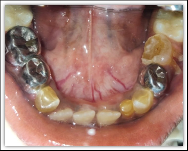
Figure 3: stainless steel crown with-74,84,85
Stage 2: Teeth with hopeless prognosis were extracted 54,55,64,65. 75 was not planned to be extracted at this stage.
Stage 3: Prosthetic rehabilitation of the upper arch with overlay denture was done. Secondary impressions were taken from special tray prepared from primary cast (Figure 4a,b). On these impressions cast was poured and trial denture was fabricated (Figure 4c). Post fabrication try in was done and adjustments according to patient’s comfort were made (Figure 4d). After making adjustments in the try in denture, final over lay denture was delivered (Figure 4e). C clasps were added bilaterally to provide added retention. Mono-plane occlusion was kept to minimize masticatory forces acting on lower teeth.
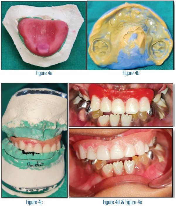
Figure 4: Prosthetic rehabilitation with overlay denture
Stage 4: Oral Hygiene instructions were enforced and patient counselling was done regarding the importance of having follow-up visits in a short period of time (for early detection of any new fracture or abscess and also to check the need for changing the prosthesis).

Figure 5a: pre-op & Figure 5b: follow-up after 3 months
DISCUSSION
Treatment was done according to AAPD guidelines.2In initial treatment molars which had severe attrition and no dehiscence were restored with stainless steel crowns (85,84,74).Wide pulp chambers and thin dentin caused difficulty in tooth preparation. Hence, stainless steel crowns were given by Hall’s technique.8 In a case report byTorija ET, stainless steel crowns were given in a 18 month old child suffering from dentinogenesis imperfecta to prevent further damage to the coronal structure.9 Other authors like Sapir S, Shapira J have also advocated early diagnosis and prevention with preformed crowns for patients suffering from dentinogenesis imperfecta.10
75 had hopeless prognosis, so stainless steel crown was not planned. Also, extraction was not planned to prevent the loss of vertical height, allow normal growth of the facial bones and temporomandibular joint and avoid interference with the eruption of the remaining permanent teeth.
Overlay dentures were planned for maxillary arch, it enhanced mastication and esthetics, maintains alveolar bone by the retention of teeth and increased proprioception. Syriac G et. al. Schneidman E et.al. also advocated rehabilitation with overlay dentures to prevent further damage.11,12 The denture along with stainless steel crowns has helped in restabilising the vertical height of the face and in turn enhance the aesthetics.
After 3 months follow up improvement in the health of the child was noted (Figure 5 a,b). This has helped him to improve his aesthetics, rebuild his self-confidence and improve the quality to his life.
CONCLUSION
Full mouth rehabilitation in patients with Dentinogenesis Imperfecta can help in improving esthetics, enhances mastication thus give a positive psychological support and better quality of life.
REFERENCES
- Talbot es. Arrests of development and decalcification of the enamel and dentine. Read in the Section of Oral and Dental Surgery, at the Forty-third Annual Meeting of the American Medical Association, held at Detroit, Mich., June, 1892. Journal of the American Medical Association. 1893 Jan 14;20(2):29-32.
- Council O. Guideline on dental management of heritable dental developmental anomalies. Pediatr Dent. 2013;35(5):179-84.
- Gupta SK, Saxena P, Jain S, Jain D. Prevalence and distribution of selected developmental dental anomalies in an Indian population. Journal of oral science. 2011;53(2):231-8.
- Takagi Y, Sasaki S. A probable common disturbance in the early stage of odontoblast differentiation in dentinogenesis imperfecta type I and type II. J Oral Pathol 1988; 17(5):208-12.
- MacDougall M, Jeffords L, Gu T, et al. Genetic linkage of the dentinogenesis imperfecta type III locus to chromosome 4q. J Dent Res 1999;78(6):1277-82.
- Witkop CJ. Hereditary defects in enamel and dentin. Acta genetica et statistica medica. 1957 Jan 1;7(1):236-9.
- Shields ED, Bixler D, El-Kafrawy AM. A proposed classification for heritable human dentine defects with a description of a new entity. Archives of oral biology. 1973 Apr 1;18(4):543-IN7.
- Page LA, Boyd DH, Davidson SE, McKay SK, Thomson WM, Innes NP. Acceptability of the Hall Technique to parents and children. NZ Dent J. 2014 Mar 1;110(1):12-7.
- Torija ET. Oral rehabilitation in dentinogenesis imperfecta. Report of a case. Revista ADM (Asociacion Dental Mexicana: 1988). 1990 Jan 1;47(1):9-11.
- Sapir S, Shapira J. Dentinogenesis imperfecta: an early treatment strategy. Pediatric dentistry. 2001 Jun;23(3):232-7.
- Syriac G, Joseph E, Rupesh S, Mathew J. Complete overlay denture for pedodontic patient with severe dentinogenesis imperfecta. International journal of clinical pediatric dentistry. 2017 Oct;10(4):394.
- Schneidman E, Wilson S, Spuller RL. Complete overlay dentures for the pediatric patient. Pediatr Dent. 1988 Sep 1;10(3):222-5.
Disclaimer from Guident
The views and opinions expressed in articles published in Guident are solely those of the authors and do not necessarily reflect the views of the editors, the editorial board or the publisher, Ivory India. Guident does not endorse any product, technique or treatment described. Clinical content is intended for qualified dental and medical professionals and is not a substitute for individual clinical judgement. Guident, its editors and the publisher accept no liability for any consequence arising from the use of information published in the journal.
How to cite this article
Dr. Khushboo Barjatya, Dr. Arushi Goel. Full Mouth Rehabilitation in Patient with Dentinogenesis Imperfecta in Primary Dentition: A Case Report. Guident. 2023 Mar;16(4). Available from: https://guident.in/articles/pedodontics/full-mouth-rehabilitation-in-patient-with-dentinogenesis-imperfecta-in-primary-dentition-a-case-report.html