Ectodermal Dysplasia
Department of Pedodontics and Preventive Dentistry K. D. Dental College, Mathura, U.P. India
INTRODUCTION
The ectodermal dysplasia includes a complex and highly diverse group of heritable disorders that share common developmental abnormalities of ectodermal derivatives. The broader definition of ectodermal dysplasia’s (as heritable disorders involving at least two of the ectodermal derivatives nails, teeth, hair, and sweat glands) encompasses 170 – 200 conditions. Some conditions included by this definition are relatively common; others are rare and in some cases, family- specific.
Ectodermal dysplasia’s represent a large and complex nosological group of congenital diseases which was first described by Thurnam in 1848 and later in the 19th century by Darwin. It is an X-linked recessive disorder. Ectodermal dysplasia was assigned to the X-chromosome in 1921 by Thadani, who later reported that carrier females could manifest signs of the condition. Their incidence is relatively rare (1/100,000 births) and is characterized by the abnormal development of the ectodermal derivative. The incidence of hereditary ectodermal dysplasia is about 0.00001%. Out of 100 affected individuals, 90 are male and females are never affected by complete syndrome. Ectodermal dysplasias are rare genetic disorders. Anhidrotic ectodermal dysplasia is inherited as X-linked recessive. Most of the cases of this syndrome are males (90%) and the females are only carriers. The complete syndrome does not occur in females; however, a much rarer form of ectodermal dysplasia inherited as autosomal recessive has been described where females present with the complete syndrome.
ETIOLOGY
- X-linked recessive HED or anhidrotic ectodermal dysplasia and immunodeficiency (EDA-ID), is caused by mutations in gene
- Autosomal dominant HED is caused by mutations in the DL gene, which encodes the EDA (ectodysplasin) receptor.
- Autosomal recessive HED may also result from mutations in the EDARADD gene, which encodes a protein that interacts with the EDA receptor.
- Hidrotic ectodermal dysplasia (Clouston syndrome), which is an autosomal dominant disorder, is caused by mutations in GJB6.
- Hay-Wells syndrome (ankyloblepharon filiforme adnatum), Rapp-Hodgkin syndrome and EEC (Ectrodactyly-ectodermal dysplasia) syndrome are all caused by mutations in the TP73L (p63) gene, featuring congenital abnormalities of skin, hair, teeth, nail, eccrine and mucous glands, as well as cleft deformities.
The genetic defects underlying several other ectodermal dysplasia’s are also known.
- Palmoplantar keratoderma with deafness is caused by mutations in the GJB26 gene, which encodes connexin 26.
- Margarita Island ectodermal dysplasia is caused by mutations in the PVRL1 gene, which encodes nectin-1.
- Ectodermal dysplasia with skin fragility is caused by mutations in the PKP1 gene, which encodes plakophilin 1.
- Hypotrichosis with juvenile macular dystrophy is caused by mutations in the CDH3 gene, which encodes p-cadherin.
- ADULT syndrome is caused by mutations in the TP63 gene.
- Split hand-foot malformation syndrome is caused by mutations in the TP63 gene.
CLASSIFICATIONS
- The most comprehensive accounts of clinical phenotypes and inheritance patterns of ectodermal dysplasia were produced by Freire-Maia and Pinheiro, 1984, in their classic monograph and in subsequent writings by Pinheiro and Freire-Maia, 1994. Freira-Maia and Pinheiro have proposed a classification based on ectodermal derivative involved, i.e. 1 to 4:
1. Indicates hair dysplasia/trichodysplasia
2. Indicates dental dysplasia
3. Indicates nail dysplasia/onychodysplasia
4. Indicates eccrine gland dysplasia/dyshidrosis.
CLINICAL MANIFESTATIONS
Clinically, hereditary ectodermal dysplasias may be divided into two broad categories:
X-linked hypohidrotic form (Christ-Siemens-Touraine syndrome), characterized by the classical triad of hypodontia, hypohidrosis and hypotrichosis and by characteristic dysmorphic facial features, presenting with the following features:
- Mostly patients show fine, sparse, lusterless, fair hair; therefore, little pigmentation in the hair shaft is observed microscopically and the medulla is often discontinuous. When medullation is present, a “bar code” appearance is often seen
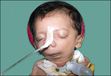
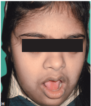
- The characteristic facies is characterized by frontal bossing, large lowest ears, wrinkled hyper- pigmented periorbital skin, thick everted lips, saddle nose and sunken cheeks. These features are not recognized until infancy.
- Dental manifestations include conical or pegged teeth, hypodontia or complete anodontia and delayed eruption of permanent teeth.
- Onychodystrophy may occur, but is not common.
- Extensive scaling of the skin and unexplained pyrexia secondary to anhidrosis may occur in the neonatal period. The development of a chronic eczematous dermatitis is common.
- Other common signs are short stature, eye abnormalities, decreased tearing and photophobia.
- Moderate built and poorly nourished.
Hidrotic form (Clouston’s syndrome) that usually spares the sweat glands but affects the teeth, hair and nails. Most of the other clinical features are similar to that seen in the hypohidrotic form. It has an autosomal-dominant inheritance and is common in persons of French-Canadian ancestry.
ORAL MANIFESTATIONS
- Aberrations in number and shape of the teeth (Figs 1 and 2 )
- Malformed teeth, i.e. conical central and lateral incisors (Fig. 3)
- Premature caries
- Gingivitis
- Early tooth loss of both the dentitions
- Premature exfoliation
- Dry and sticky oral mucosa
- Knife-edge alveolar ridges (Fig. 4 )
- Partial anodontia/hypodontia (Figs 5 and 6 ).
RADIOLOGICAL MANIFESTATIONS
- Altered morphology of the teeth
- Aplasia of alveolar bone
- Mandibular aplasia
- Decreased facial height involving lower one-third of the face (Fig 7).

Figs 1-6: Intraoral manifestations—aberrations in number and shape, conical central and lateral incisors, dry sticky mucosa and knife-edge alveolar ridges

Fig 7: OPG showing partial anodontia of upper jaw and complete anodontia of lower jaw along with aplasia of bones
MANAGEMENT
Medical management is symptomatic. Patients with hypohidrosis require frequent consumption of cool liquids, wear cool clothing and should stay in a cool environment (air conditioning). Emollients for dry skin, artificial tears for patients with decreased lacrimation, saline sprays for nasal mucosa and antibiotics, if any infection occurs, may be required. Dental malalignment may require orthodontic treatment.
Surgery is required mainly for oral and dental rehabilitation. Cleft lip or palate, if present, requires surgical repair. Other midfacial defects or hand/foot deformities may be corrected surgically. The oral rehabilitation of patients presenting with congenitally missing dentition is challenging because of the need for a multidisciplinary approach. However, prosthesis is certainly the answer for such patients. Additional considerations, such as the patient’s age, stage of growth, inherent anatomic deficiencies present in conjunction with the missing teeth, soft tissue defects, existence of malformed dentition, severe diastemas and psychological status, must be considered. Patients with ectodermal dysplasia because of tooth absence have reduced alveolar bone with “knife-edge” morphology, making implant reconstruction a challenge.
Disclaimer from Guident
The views and opinions expressed in articles published in Guident are solely those of the authors and do not necessarily reflect the views of the editors, the editorial board or the publisher, Ivory India. Guident does not endorse any product, technique or treatment described. Clinical content is intended for qualified dental and medical professionals and is not a substitute for individual clinical judgement. Guident, its editors and the publisher accept no liability for any consequence arising from the use of information published in the journal.
How to cite this article
Dr. Priya Verma. Ectodermal Dysplasia. Guident. 2018 Nov;11(12). Available from: https://guident.in/articles/pedodontics/ectodermal-dysplasia.html