Surgical Guides in Dental Implants: A Review
Department of Prosthodontics and Crown & Bridge, K.D. Dental College & Hospital, Mathura, UP, India.
ABSTRACT
The use of dental implants in recent years has grown quickly with the advent of osseointegration. Inadequate diagnosis, treatment planning, surgical technique, and implant placement frequently result in unintended implant problems. By employing surgical guidance for implant location, this can be avoided. Although few practitioners employ it, guided surgery is acknowledged as the most accurate approach to put an implant and predictably relate the implant to its final prosthesis. A prosthetically driven implant prosthesis ensures good looks, functionality, and most importantly, hygienic upkeep for long-term success. The use of surgical templates has made it possible for clinicians to provide predictable surgical and prosthetic rehabilitation. In addition to reducing the risk of iatrogenic injury to important anatomical structures, surgical guides have significantly increased the aesthetic and functional benefits of prosthodontic-driven implant surgery. The authors provide an overview of the usage of surgical guides in clinical situations within the confines of this paper.
Keywords: Pilot guides, Circumference lead strip guides, Gutta-percha guides
INTRODUCTION
Dental implants are now a popular therapeutic option for replacing missing teeth. The introduction of dental implants has significantly impacted patients, and implant-supported oral restorations are now a common treatment option for patients with partial or complete dentures, severe bone loss, and locations that were previously thought to be unsuitable for implant placement. These procedures involve bone augmentation, regeneration, and soft tissue regeneration. The use of medical imaging and software planning has led to considerable improvement in treatment planning. Surgical guides now play an important role in transferring this premapped plan to placing the implants at their designated positions (angulation and depth).1 More precise information is needed when the host bone is weakened or there is a higher desire for aesthetics, which justifies the use of more complex design approaches. Recently, emphasis has shifted from free handed implant placement techniques in adequate available host bone (assessed by the surgeon at the time of surgery) to placing implants with nearly exact prediction of the final surgical/prosthetic outcome by means of computer-guided surgical systems.2 To facilitate accurate translation from the desired surgical treatment plan to reality, templates or surgical guides can be used.3 Systems that can help the clinician with diagnosis, treatment planning, and the surgical procedure itself have been developed thanks to advancements in computer technology (hardware/software). The precision and dependability of three-dimensional computer-assisted interactive implant planning software tools are sufficient for reliable clinical application. Direct navigation and stereo lithographic drill guides are two technologies for computer-based transmission that are available. Through guided drilling templates, the latter enables a trustworthy transfer of the surgical plan to the surgical field, assisting the surgeon in correctly positioning dental implants while fully anticipating the final prosthetic outcome, soft tissue management, emergence profile, and tooth morphology. Using computer technology also aids in patient satisfaction because surgical times are shorter, treatment can be less invasive, healing times can be shorter, and there are less chances of clinical complications.4,5
CLASSIFICATION OF SURGICAL GUIDE
(A): Based on the area of operation
Guides for partially edentulous sites (tooth supported or bone supported-depending on amount of the edentulous space) and Guides for completely edentulous sites (mucosa or bone supported).1
(B): Based on the support the surgical guides derive
Tooth supported, Bone supported, and Mucosa supported. Rules for planning usage of different types of surgical guides.6
- Tooth supported guides: Minimum three stable teeth should be present to support the guide during surgery [Figure 1]
- Mucosa supported guides: It is used in fully edentulous sites. Advantage-less or no tissue reflection is required, so less postoperative discomfort. Will require scan prosthesis and surgical guides during surgery [Figure 2]
- Bone supported guides: They are used in partially edentulous sites and completely edentulous sites. When used in partially edentulous sites, it should possess at least 3 cm of supporting bone or 3 teeth would need replacement. Bone guides are especially used when edentulous sites possess thin bone. Raised flap should provide a good view to implant sites and insertion of guides [Figure 3]

Figure 1: Supported surgical

Figure 2: Mucosa supported surgical guide

Figure 3: Bone supported Surgical guide
(C): Based on the accessibility
Surgical guides can be open sleeve or closed sleeve (increased accessibility).6
(D): Based on Utility
- Pilot guides: The sleeves only allow pilot drills. Angulation control is achieved. Depth control is to be obtained manually by assessing markings on drills. Later, the surgical guide is removed, and osteotomy site is expanded in the absence of surgical guide
- Complete drill guides: It uses drill keys or sleeves. Different sleeves for different diameters of drills, which are changed concomitantly as osteotomy is widened. Angulation as well size of osteotomy is controlled by guide; depth is controlled manually
- Safe guides/easy guides: Uses drill key or sleeves as above with additional implant stopper that controls the depth of drilling. Allows both osteotomy preparations with surgical drills and installation of implants.6,7
(E): Based on material
Self/light cure acrylic resin, metal reinforced acrylic templates; vacuum formed polymers, milling, CAD-CAM prosthesis, stereo lithographic models. The surgical accuracy of manually processed resin and vacuum formed guides is less in comparison to the latter mentioned milling, CAD-CAM prosthesis or stereo lithographic models.6,8
FABRICATION PROCESS OF SURGICAL GUIDES 9 :
Four most commonly used surgical guides are discussed in this article
A. Circumference lead strip guides
- Pour diagnostic casts in stone.
- Complete a diagnostic wax-up using hand articulation or a precision articulator. This establishes the desired prosthetic orientation of each tooth inthe edentulous area.
- Use wax to block out all undercuts on at least 2 teeth on either side of the edentulous area. Coat the lingual/palatal, buccal, and occlusal surfaces of the cast with a lubricant.
- Make a template using a 0.020-in., vacuum-formed, thermoplastic material such as Thermoforming Material (T&S Dental & Plastics Mfg, Myerstown,Pa.).
- Separate the vacuum-formed shell template from the cast, and trim it
- Remove the wax-up from the cast. Reline the vacuum-formed template in the edentulous area with custom tray material (Triad, Dentsply Co, York, Pa.) or autopolymerizing resin (Jet Acrylic, Lang Dental Mfg Co, Wheeling, Ill.), and set it back on the cast. Filling the shell template with resin down to the edentulous part of the cast will strengthen the template.
- For enhanced stability in the mouth, make sure that the template extends at least 2 teeth on either side of the edentulous area. When the site is a distal abutment, extend the template anteriorly and, if necessary, across the arch and past the canine.
- Make a prosthetic determination of the most appropriate placement of the radiopaque indicator for each surgical site. Create a circumference radiopaque indicator by gluing a 2-mm wide lead strip, cut from any intraoral dental film lead sheet, to the buccal, occlusal, and lingual/palatal surfaces of the template, outlining the tooth over the implant site (Figure 4).

Figure 4: Demonstration model with circumference guide in area of maxillary left first molar
B. Vertical lead strip guides
- Complete steps 1 through 6 outlined in the circumference guide fabrication section.
- Separate the vacuum-formed template from the resin tooth. Connect the resin tooth by using a layer of resin placed on the occlusal and lingual/palatal surfaces of at least 2 teeth on either side of the edentulous area. When the site is a distal abutment, extend the template anteriorly and, if necessary, across the arch and past the canine
- Make a prosthetic determination of the most appropriate placement of the access hole for each surgical site. Drill buccal access grooves in the template over the planned prostheticimplant sites. Create a vertical radiopaque indicator from a 2-mm wide lead strip cut from any intraoral dental film lead sheet, and vertically glue it on the lingual/palatal wall of the groove (Figure 5)

Figure 5: Demonstration model with lead strip guide in area of mandibular right first and second
C. Gutta-percha guides
- Complete steps 1 through 6 outlined in the circumference guide fabrication section.
- Follow with step 2 outlined in the vertical lead strip guide fabrication section.
- Make a prosthetic determination of the most appropriate placement of the access hole for each surgical site. Use a 5⁄32-in. drill bit in a laboratory handpiece to drill an access hole through the template over the planned prosthetic implant site. A surveyor may be used as an aid to determine the inclination of the access hole. Condense guttapercha (Hygenic, The Hygenic Co, Akron, Ohio) to fill the access hole (Figure 6)
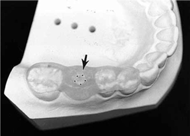
Figure 6: Demonstration model with gutta-percha guide in area of mandibular right first molar
D. Metal sleeve guides
- Complete steps 1 and 2 outlined in the circumference guide fabrication section.
- Make a prosthetic determination of the most appropriate angulation and placement of the access holes for each site. Use a 3⁄32-in. drill bit in a laboratory hand piece to drill through the diagnostic wax-up into the stone cast to a depth of at least 10 mm. This hole in the cast represents the osteotomy site in the alveolar ridge
- Remove the wax-up from the cast. Insert the smallest end of an 18-mm Guide Right (DePlaque Corp, Victor, N.Y.) pin into the 3⁄32-in. hole prepared in the cast, providing the orientation that needs to be captured and transferred to the guide sleeve. Place an appropriate Guide Right stainless steel sleeve over the pin. The sleeves are precision cut and deburred electrochemically, and they come in multiple diameters and lengths
- Use wax to block out all undercuts on at least 2 teeth on either side of the edentulous area. Coat the lingual/palatal, buccal, and occlusal surfaces of the cast with a lubricant
- Secure the relationship between the sleeve and adjacent teeth with custom tray material (Triad, Dentsply Co) or autopolymerizing resin (Jet Acrylic, Lang Dental Mfg Co) as the base material. Extend the template with a layer of resin placed on the occlusal and lingual/palatal surfaces of at least 2 teeth on either side of the edentulous area (Figure 7).

Figure 7: Demonstration model with metal sleeve guide in area of maxillary left lateral incisor
USES OF SURGICAL GUIDES
- Osteotomy drills are guided to the proper depth, angulation, and location.
- Guidance of implant fixtures at the appropriate depth, angulation, and position
- Providing recommendations for the quantity of hard and soft tissues to be harvested, as well as any necessary bone reduction.
ADVANTAGES
- Reduces the chance of human mistake during free-hand implant implantation
- Minimally invasive procedure—since surgical guides require less involvement, postoperative surgical issues are reduced, benefiting both the patient and the doctor psychologically.
- Accuracy – Because implants are prosthetically driven parts, any error might cause a sudden change in how they work. Implant placement has improved with surgical guidance.
- The most important consideration when placing implants in important mouth locations is safety. The smallest mistake might have serious consequences.
- Such deviations can be avoided with guidelines. Damage to vital structures can be easily avoided
- Predictability It is impossible to remain alert throughout the entire procedure. When compared to guided implantation procedure, even skilled hands produce inferior results.
- The use of surgical guidance to translate software planning to the real placement has demonstrated good cosmetic outcomes in the areas of aesthetics and hygiene. Correct implant placement ensures maintenance of good oral health.
- Implant supported prostheses should be positioned in predetermined locations for the best chance of survival. Guides can assist with such accurate implant placement.
- Shortening the duration of implant operation • Ease of fabrication: Most implants have built-in software that makes it simple to purchase and shop online with the click of a button.
DISCUSSION
In the manner we assess the problem and arrange the course of therapy, digital technology has proven to be a useful aid. However, if a strategy is poorly carried out, even the finest of intentions seem useless. Guided surgery can be used with or without elevating a full-thickness flap. In this patient, a flapless approach was chosen because of the availability of adequate keratinized tissue and bone volume that would require no contouring or other grafting procedures.10 During oral implant placement, the drill (position, depth and angulation) must be guided by the surgeon according to the final form of the prosthetics. Ideal placement facilitates the establishment of favorable forces on the implants and the prosthetic component. In this regard, surgical guides have shown better predictability of placement improving better prosthetic results.11 Numerous guides, including self/light cure acrylic resin, metal reinforced acrylic templates, vacuum formed polymers, milling, CAD/CAM prosthesis, and stereolithographic models, have been published in the literature. The guides and disks discussed in this article are tools that are suggested for use with cross-sectional tomography during the pre-implant evaluation of surgical sites. They should assist the implant team in transferring important imaging data from the prosthetic and diagnostic phases to the surgical stage. This entails understanding the relationships between the planned prosthetic angulation and the residual bone angulation, as well as the relationships between the planned implant angulation and anatomical structures and nearby implants.
CONCLUSION
Digital dentistry is being used as a “Two-edge-sword”. The standard of care in many complicated dental circumstances has been recommended to be virtual implant design and guided surgery, which is growing in popularity. When location is crucial due to anatomical conditions, a precise surgical template is especially helpful. Avoiding essential structures, placing the implant in a narrow maxilla, and avoiding fenestration when the cortical plates are parallel all help. It eliminates the need to insert shorter or smaller diameter implants than planned in order to give a necessary margin of safety, and it permits placing an implant where manual placement would result in failure when bone anatomy necessitates very accurate placement. Within the confines of the article the author have discussed the types, fabrication and uses of the commonly used surgical guides.
REFERENCES
- T Umapathy, Chirenjeevi Jayam, BS Anila, CP Ashwini.Overview of surgical guides for implant therapy. Journal of Dental Implants. Jan - Jun 2015.5(1)
- GanzSD.“Whatis the single most important aspect of implant dentistry”- Implant Soc 1994;5(1):2–4.
- Ganz SD. Presurgical planning with CT-derived fabrication of surgical guides. J Oral Maxillofac Surg 2005;63(9 Suppl 2):59–71.
- Arisan V, Bolukbasi N, Oksuz L. Computer-assisted flapless implant placement reduces the incidence of surgery-related bacteremia. Clin Oral Investig 2012; 17(9):1985–93.
- Sicilia A, Botticelli D. Computer-guided implant therapy and soft- and hard-tissue aspects. The Third EAO Consensus Conference 2012. Clin Oral Implants Res 2012;23(Suppl 6):157–61.
- Drill guides for every case scenario: Surgi Guide Cookbook. Available from: http://www.materialisedental.com/materialise/view/en/2395185.SurgiGuide+dental+drill+guide+Cookbook.html. [Last cited on 2013 Aug 25].
- Zitzmann NU, Marinello CP. Treatment plan for restoring the edentulous maxilla with implant-supported restorations: Removable overdenture versus fixed partial denture design. J Prosthet Dent 1999;82:188-96
- Fortin T, Champleboux G, Lormée J, Coudert JL. Precise dental implant placement in bone using surgical guides in conjunction with medical imaging techniques. J Oral Implantol 2000;26:300-3
- Almog.D.V, Torrado.E, Meitner.S.W. Fabrication of imaging and surgical guides for dental implants. The Journal Of Prosthetic Dentistry. 2001;85:504-8.
- da Rosa EL, Oleskovicz CF, Aragao BN. Rapid prototyping in maxillofacial surgery and traumatology: case report. Braz Dent J 2004;15:243-7.
- Orentlicher G, Abboud M. Guided surgery for implant therapy. Oral Maxillofac Surg Clin North Am 2011;23:239-56
Disclaimer from Guident
The views and opinions expressed in articles published in Guident are solely those of the authors and do not necessarily reflect the views of the editors, the editorial board or the publisher, Ivory India. Guident does not endorse any product, technique or treatment described. Clinical content is intended for qualified dental and medical professionals and is not a substitute for individual clinical judgement. Guident, its editors and the publisher accept no liability for any consequence arising from the use of information published in the journal.
How to cite this article
Dr. Vishal Paul, Dr. Manesh Lahori, Dr. Siddharth Sisodia. Surgical Guides in Dental Implants: A Review. Guident. 2023 Jul;16(8). Available from: https://guident.in/articles/implantology/surgical-guides-in-dental-implants-a-review.html