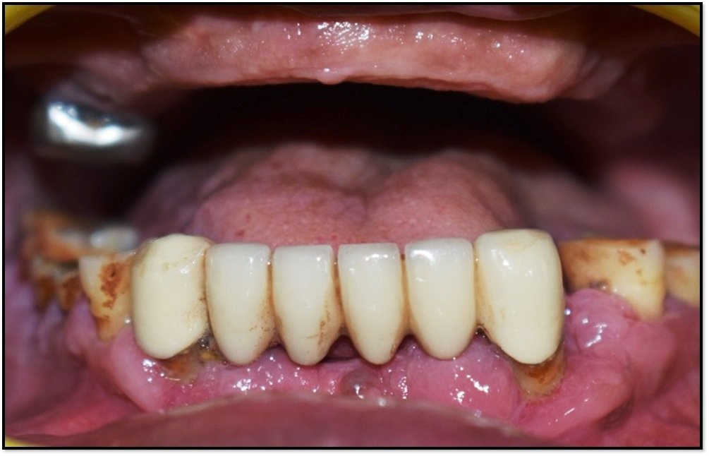
Rehabilitation of Partially Edentulous Mandibular Posterior Region with Implant-Retained Prosthesis-a Case Report
Department of Prosthodontics, Post Graduate Institute of Dental Sciences, Rohtak, Har., India.
ABSTRACT
Teeth are designed for lifetime but some patients lose their teeth partially or completely before time due to several causes like dental caries, periodontal problems, traumatic fractures of teeth and so on. Replacing missing teeth is important for patient’s oral as well as general health. With advancement of dental science there are various treatment options available to rehabilitate a patient with several missing teeth that to functionally and aesthetically sound. Public awareness about aesthetics makes it more challenging for dental practitioner to provide the best treatment with acceptable outcome. Implant treatment is considered as a good treatment option over conventional treatment options like fixed bridge prosthesis and removable partial or complete dentures. This case report discusses the prosthetic rehabilitation of missing mandibular posterior tooth with implant supported prosthesis.
Keywords: Dental implants; Implant-retained prosthesis; Partially edentulous jaws; Screw retained; Provisional restoration.
INTRODUCTION
The goal of modern dentistry is to achieve aesthetically and functionally sound stomatognathic system.1 However, in patient having several missing teeth, it becomes difficult to achieve the goal. In partial edentulism, implant-borne rehabilitation is a good treatment option for such patients.2 Dental implant is a prosthesis that forms connection with bone of jaw or skull to support a dental prosthesis. A highly biocompatible, pure titanium alloy is used as a implant material. Dental implant alleviates the quality of treatment and life for the patients with multiple missing tooth.3 The success rate of implant varies case to case depending upon several factors like bone density, bone volume, soft tissues, force factors, treatment planning and skill of the implant surgeon.4
CASE 1
A 51-year-old female patient reported to the Department of Prosthodontics, with the chief complaint of missing tooth in lower left back tooth region for past 1year. The patient had full compliment of teeth in both arches except left mandibular second premolar. The remaining tooth were in good condition and periodontally sound with two teeth having amalgam filling. Patient wanted replacement of missing tooth for aesthetic purpose. On intraoral examination, mild resorbed ridge in missing tooth region seen. Additionally, adequate bone height and mesio-distal and bucco-lingual width were present for fixed treatment. In the view of clinical examination and all treatment options discussed and patient opted for the conservative treatment with respect to missing left mandibular second premolar region.
CASE TREATMENT
A. Pre-operative Investigation
An intraoral periapical (IOPA) radiograph with respect to left mandibular second premolar region was advised (Fig. 1). The bucco-lingual and mesio-distal width and height of bone were measured in the radiograph. Irreversible hydrocolloid impression material (Algitex Alginate Impression Material-Dental Product of India) was used for making maxillary and mandibular diagnostic impression. The dental stone model was obtained (Kalabhai dental stone-Dental product of India) for measuring the bucco-lingual and mesio-distal width of missing tooth region clinically. The radiographic values and clinical values were compared. Cone Beam Computed Tomography (CBCT) scan was also done for detailed information in 3-dimensional view. The 3D imaging software (Carestream Dental-product of United States of America) was used to analyse the bone density and dimension of missing tooth region and also for virtual placement of implant with respect to left mandibular second premolar region along with distance of implant from vital structures like mental foramen, mandibular canal and mandibular nerve, distance of implant from adjacent teeth and buccal and lingual cortical plate. Complete Hemogram, Random blood sugar test and Blood pressure test were conducted.
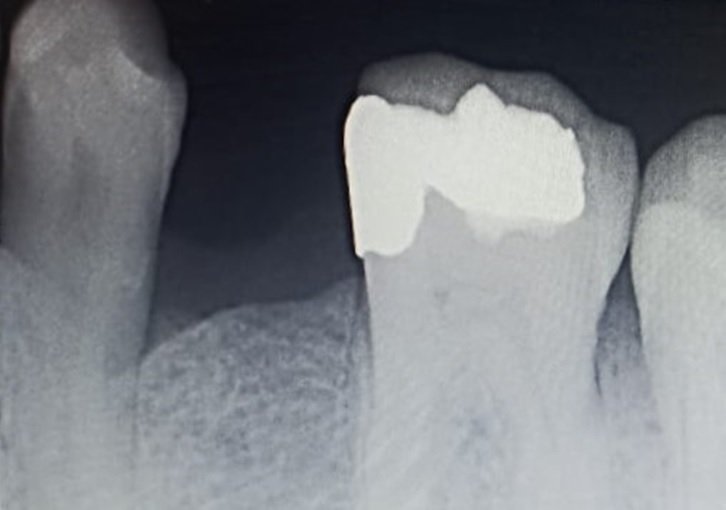
Fig. 1: Preoperative radiograph
B. Intra-operative procedure
Ask patient to rinse with 0.12% chlorhexidine (Periogard) to reduce the bacterial load of oral cavity and post-implantation complications. 1.5 ml local anaesthetic 2% Lignocaine hydrochloride with adrenaline 1:80,000 was administered for Inferior alveolar nerve block and 0.5 ml injected for Lingual nerve block. After 5 minutes area of surgical concern was checked for objective and subjective signs. The horizontal mid crestal incision was placed with respect to left mandibular second premolar region with surgical blade no. 15 and crevicular incision was placed with respect to adjacent teeth as releasing incision with surgical blade no. 12 (Fig. 2) Facial and lingual mucoperiosteal flaps elevated with Periosteal elevator to expose the ridge crest (Fig. 3).
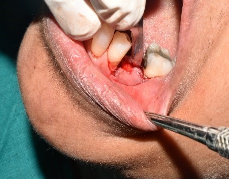
Fig. 2: Placement of incision
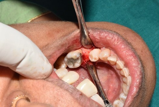
Fig. 3: Reflection of mucoperiosteal flaps
a. Pilot drilling: After exposing the underlying bony ridge, the reference of adjacent tooth was taken to prepare implant osteotomy site. A small round bur was used to mark the implant site and drill through high density crestal bone. A sharp pointed lance drill or pilot drill was used as first drill to make initial entry into the bone (Fig. 4). Drilling was done at speed of 1200-2500 rpm under a constant stream of chilled saline with torque adjusted between 30 and 50 Ncm.
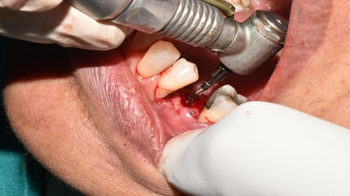
Fig. 4: Pilot drilling up to planned depth
b. Depth drilling: The depth drilling was done which was 2mm of diameter first used to the partial depth and direction of drilling is 3-dimensionally was evaluated by inserting parallel pin in partially prepared osteotomy. After evaluation of direction, depth drill was further used to complete the osteotomy depth.
c. Osteotomy widening: After Pilot drilling to the planned depth, osteotomy widening drills were used sequentially to the same depth. Final drill was approximately 0.4 mm less than diameter of implant placed (Fig. 5). In this case sequential drilling was done until 3.65 mm according to the size of implant used. After the final drill, prepared osteotomy was checked using depth probe.
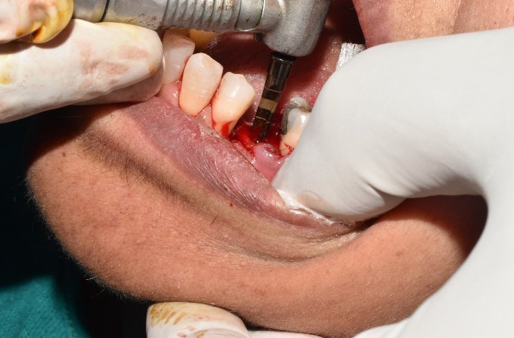
Fig. 5: Final drill
d. Implant placement: The implant (SuperLine Dentium implant system-product of United States of America) 8mm in length and 4mm in diameter was used. Implant driver was engaged into the implant connection to remove the implant from sterile vial. The implant was carried to prepared osteotomy and screwed in with torque of 35 Nm using hand ratchet (Fig. 6,7). After screwing in implant up to crest level healing abutment was placed. The simple interrupted suture with 3-0 silk suture reverse cutting (Perma Hand Ethicon) were placed to approximate the flaps with a tension free primary closure (Fig. 8). An intraoral periapical radiograph was done for the respective site of surgery (Fig. 9).

Fig. 6 and 7:Placement of implant fixture and torque
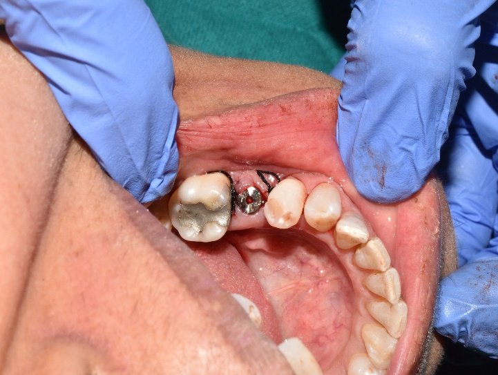
Fig. 8: Placement of healing abutment and primary closure with suture
POST-OPERATIVE CARE
Post-operatively patient was instructed for maintaining good oral hygiene, warm water mouth rinses 3 times a day. Postoperative medications was advised as per need. After 1 week patient was recalled for suture removal. Patient was recalled for regular follow up for assessment of surgical site.
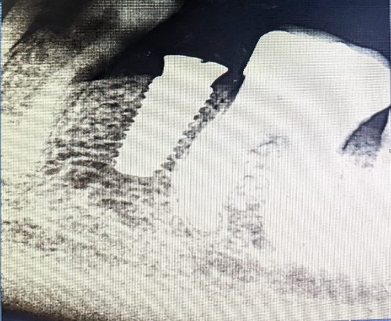
Fig. 9: Post-operative intraoral radiograph
PROSTHETIC PHASE
After 4 months patient was recalled and maxillary and mandibular impressions were made with addition silicone (Orikam Neopure- A & C Silicone impression materials). Cast was made using Type III gypsum (Kalabhai dental stone-Dental product of India). All-ceramic crown prosthesis was fabricated, cementation of prosthesis was done with respect to missing left second premolar region (Fig. 10).
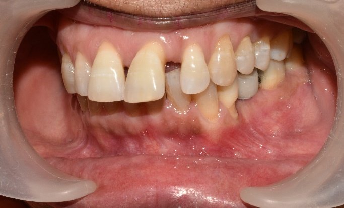
Fig. 10: Placement of implant-retained prosthesis with respect to missing left second premolar region
DISCUSSION
Fixed dental prosthesis is considered as a good treatment option over removable prosthesis for replacement of missing teeth. Removable prosthesis lacks in providing retention and functional stability, also cause discomfort to patient.5 Fixed dental treatment is of two types: tooth-supported prosthesis or implant-retained prosthesis.
Tooth supported prosthesis involves unnecessary alteration of natural teeth, intentional root canal treatment also supports from adjacent tooth affect the abutment tooth because of the masticatory forces occurring on the of 3-unit prosthesis.6 The occlusal load more than adaptive range on the 3-unit prosthesis distributes to the abutment teeth which may cause periodontal weaking or failure of prosthesis.
On the other hand implant-retained prosthesis seems to be a better option as there is preservation of natural structure.7 In recent years, implant treatment planning has changed from surgical approach to prosthesis-oriented planning with 3D imaging and CAD software. It requires accurate treatment planning for implant placement.8,9 Implant surgery involves several steps, in which if proper sterilization measures are not followed it will lead to post-implantation infection complications. This advent digital technology has remarkably improved the quality of treatment provided to the patient. With CT-guided dental implant surgery, better clinical experience and improved outcomes can be provided to patients with confidence.10 The pre-operative digital planning helps in choosing the implant length and diameter that fit properly in the biologic boundaries. Assessment of the depth of first drill and alignment of drill are very crucial steps in osteotomy preparation. Additionally, distance of virtually placed implant from vital structures can be assessed.11
As implant treatment is an invasive surgical treatment and if not performed properly can lead to severe medical emergencies. Success of implant treatment depends on the skill of the implant surgeon and oral hygiene maintenance by the patient. Otherwise, implant-retained prosthesis is a good treatment option as improves the quality of life and comfort of the patient.12
CONCLUSION
Implant treatment is considered as a good treatment option for replacement of missing teeth, without taking support from adjacent tooth. One of the best advantage is, there is no need of unnecessary preparation of natural tooth structure adjacent to the missing space to be used as abutment tooth. Good surgical and prosthetic protocols with proper case planning on the part of practitioner and meticulous post-operative care and regular oral hygiene maintenance by the patients are keys for a successful outcome.
REFERENCES
- Priest G. A Current Perspective on Screw-Retained Single-Implant Restorations: A Review of Pertinent Literature. J Esthet Restor Dent. 2017;29(3):161-171.
- Wolfart S, Rittich A, Groß K, Hartkamp O, von der Stück A, Raith S et al. Cemented versus screw-retained posterior implant-supported single crowns: A 24-month randomized controlled clinical trial. Clin Oral Implants Res. 2021;32(12):1484-1495.
- Monteiro DR, Silva EV, Pellizzer EP, Filho OM, Goiato MC. Posterior partially edentulous jaws, planning a rehabilitation with dental implants. World J Clin Cases. 2015;3(1):65-76.
- Fortin T, Isidori M, Bouchet H. Placement of posterior maxillary implants in partially edentulous patients with severe bone deficiency using CAD/CAM guidance to avoid sinus grafting: a clinical report of procedure. Int J Oral Maxillofac Implants. 2009;24(1):96-102.
- Hölken F, Al-Nawas B, Meereis M, Bjelopavlovic M. Digital Workflow for Implant Placement and Immediate Chairside Provisionalization of a Novel Implant System without Abutment-A Case Report. Medicina (Kaunas). 2022;58(11):1612.
- Krekmanov L. Placement of posterior mandibular and maxillary implants in patients with severe bone deficiency: a clinical report of procedure. Int J Oral Maxillofac Implants. 2000;15(5):722-30.
- Rathee M, Ahlawat A, Chhikara N, Divakar S, Chahal S. Prosthodontic Rehabilitation of Young Adult Ectodermal Dysplasia Patient with Immediately Loaded Basal implants: A Case Report. 2022;26(1):3496-3504.
- Ahlawat A, Rathee M, Divakar S, Singh S Chahal S, Wakure P. Implant-Supported Hybrid Prosthesis using All on Six Concept for Atrophic Ridges with Severe Gag Reflex: A Case Report. JPR. 2022;34(16A):8-14.
- Nishimura RD, Beumer J 3rd, Perri GR, Davodi A. Implants in the partially edentulous patient: restorative considerations. J Calif Dent Assoc. 1997;25(12):866-71.
- Rangert BR, Sullivan RM, Jemt TM. Load factor control for implants in the posterior partially edentulous segment. Int J Oral Maxillofac Implants. 1997;12(3):360-70.
- Madani AS, Moeintaghavi A, Rezaeei M. Occlusal rehabilitation in a partially edentulous patient with lost vertical dimension using dental implants: a clinical report. J Contemp Dent Pract. 2010;11(6):E058-64.
- Madani AS, Moeintaghavi A, Rezaeei M. Occlusal rehabilitation in a partially edentulous patient with lost vertical dimension using dental implants: a clinical report. J Contemp Dent Pract. 2010;11(6):E058-64.
Disclaimer from Guident
The views and opinions expressed in articles published in Guident are solely those of the authors and do not necessarily reflect the views of the editors, the editorial board or the publisher, Ivory India. Guident does not endorse any product, technique or treatment described. Clinical content is intended for qualified dental and medical professionals and is not a substitute for individual clinical judgement. Guident, its editors and the publisher accept no liability for any consequence arising from the use of information published in the journal.
How to cite this article
Dr. Manu Rathee, Dr. Kritika Diwan, Dr. Sarthak Singh Tomar. Rehabilitation of Partially Edentulous Mandibular Posterior Region with Implant-Retained Prosthesis-a Case Report. Guident. 2023 Jul;16(8). Available from: https://guident.in/articles/implantology/rehabilitation-of-partially-edentulous-mandibular-posterior-region-with-implant-retained-prosthesis-a-case.html