Methods of the Dental Implant Surface Modification
Department of Faculty Dental Surgery and Implantology. Russia
Most part of the implants produced around the world are made of titanium and its alloys. It is actual not only in the relation of dental implants but also implants which replace mineralized tissues of the other skeleton areas. Titanium alloys, essentially titanium-aluminum- palladium alloy (also called Grade 5) can have improved strength properties, but because of the toxic admixtures their use in the present time is restricted in favor of the 4 types of commercially pure titanium (Steinemann S., 2000). Regardless of the presence of the bigger amount of alloying components in its composition, vast majority of implants produced in the world are made of commercially pure titanium Grade 4, having better physical and mechanicalparameters. (Table 1).
In case of contact with oxygen or tissue liquids this high reactive group of metals is able to form the stable oxide layer of 4-5 nm in depth on their surface literally in nanoseconds. This effect is undoubtedly favorable for dental implants functioning, as it minimizes the bio-corrosive processes in the implants thenselves. It is well known that in case of deep implant insertion into the bone bed there is the wear and the damage of the oxide layer, but in this case it is easily formed again; this process got the name of the surface repassivation.
Quality and quantity factors of osteo-integration depend directly on the implant surface topography as well as its chemical composition. The technological efforts related to the optimization of the implant surface must be grounded on the science-based prerequisites, based on the clear understanding of the fact which implant surface microstructure parameters are able to influence the osteo-integrative processes. It is appropriate to dwell on the issue of these factors here.
HYDROPHYLIC PROPERTIES OF THE SURFACE.
This factor has a great influence on the osteo-integration. The hydrophilic surface is more preferable if we compare it to the hydrophobic, as here the increased ability of the implant surface to interact with biological fluids and cellular elements is observed, which is considered to be of great importance at the early stages of the osteo-integration process. (Buser D, et al., 2004; Zhao G et al., 2005) The hydrophilic properties rate is defined by the method of contact angle (wetting angle) measurement and ranges from 0Ëš (that means hydrophilic) up to 140Ëš (standing for hydrophobic one) (Buser D. et al., 2004).Recent research carried out on laboratory animals showed that hydrophilic surface of the Straumann SLA implants contributes a lot to the increase of the total number of contacts on the implant and bone tissue boarder if compared to the ordinary SLA surface (Buser D, et al., 2004; Bagno A, et al., 2004; Giavaresi G, et al., 2003). Nevertheless all the
|
Element |
Grade 1, % |
Grade 2, % |
Grade 3, % |
Grade 4, % |
Ti-6Al-4V, % |
|
Nitrogen |
0,03 |
0,03 |
0,05 |
0,05 |
-0,05 |
|
Carbon |
0,1 |
0,1 |
0,1 |
0,1 |
-0,1 |
|
Hydrogen |
0,015 |
0,015 |
0,015 |
0,015 |
-0,015 |
|
Iron |
0,2 |
0,3 |
0,3 |
0,5 |
-0,4 |
|
Oxygen |
0,18 |
0,25 |
0,35 |
0,5 |
-0,2 |
|
Aluminum |
no |
no |
no |
No |
(5,5-6,75) |
|
Vanadium |
no |
no |
no |
No |
(3,5-4,5) |
|
Titanium |
rest |
rest |
rest |
Rest |
rest |
Table 1.Chemical composition of titanium according to ISO 5832/II and ASTM F 67-89
previous research carried out in collaboration with Albrektsson didn’t show any important difference between osteo-integration rates of hydrophilic surface implants and hydrophobic surface implants. (Carlsson L et al., 1989; Wennerberg A et al., 1991).
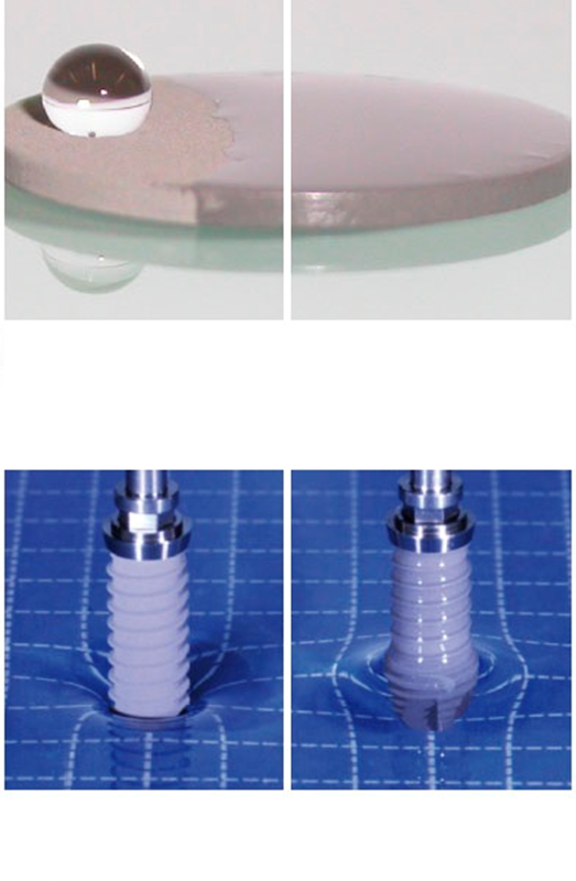
Hydrophylic properties of the surface
B. SURFACE ROUGHNESS
Depending on its evidence having a wide range of variations, the above-mentioned factor can be subdivided into macro-, micro- and nano-roughness categories. (Cochran DL et al., 1998; Wennerberg A et al., 1998).
The macroscopic roughness level is characterized by the size of the surface irregularities ranging from 1 mm to the microinch dimensions. The range of research works demonstrates the fact that the high surface roughness rate produces a positive effect on the implant mechanical stability as in the moment of its placement as in the specific terms of its functioning. (Buser D et al., 1991; Gotfredsen K et al., 1995; Wennerberg A et al., 1996) Considerable implant surface relief favors the fact that micro-irregularities of the implant surface provide a kind of interlocking between the newly formed bone tissue and implant surface itself. But you should keep in consideration the fact that considerable roughness creates prerequisites for microorganism adhesion around implants, as well as prerequisites for the peri-implantitis (peri-implant pathology) development, as well as favors the metal ions and their combinations drain and their diffusion into surrounding tissues (Becker W et al., 2000). Moderately considerable roughness of 1-2 microns in size is able to level these two undesirable phenomena. (Albrektsson T. et al., 2005).
The microscopic roughness rate reflects the micro-geometry of implant surface with irregularities ranging from 1 to 10 microns in size. This range provides the maximum rate of the implant and mineralized bone tissue interlocking. Experimental studies denote the fact that the surface relief characterized by the presence of round pores of about 1.5 microns in depth and of 4 microns in diameter can be considered as optimal ones for osteo-integration. (Hansson S et al., 1999) Implants with considerable surface micro-relief acquire greater advantages in clinical practice in conditions of the compromised state of the implant bed, which is observed in case of systemic osteoporosis and other metabolic disorders of mineralized tissues. According to the data provided by Testori T and Conner et al., the placement of short implants with considerable micro-relief in case of jaw substance deficiency has better clinical results if compared to the smooth implants of the same length. (Testori T. et al., 2001; Conner K. et al., 2003) The series of the scientific publications proves the fact of the presence of maximal amount of contacts between the bone bed and endosteal implant part with the roughness characteristics specified. Implants with above-mentioned roughness characteristics show the most resistance while pulling-out test which can also be considered as the advantage. Clinical data don’t look so encouraging, though. Esposito et al. didn’t note any statistically significant difference between the implants with different surface types in their clinical usage. (Esposito M et al., 2005).
The organization of surface on the nano-level plays an important role in the regulation of processes of biological substances adsorption on the implant surface, for example the adsorption of fibronectin, the components of the damaged cells and blood, which can subsequently influence the intensity of migration, differentiation and proliferation of the osteogenic cells (Brett P.M. et al., 2004) Unfortunately, the ways of chemical processing of implant surface for controlled formation of the surface with the necessary nano-level three-dimensional organization still stay imperfect. The problem of the choice of surface with optimal nano-level topology in the context of intensification of the osteoblastic cell adhesion on the implant surface also still has no ready-made solution.
The wide range of ways for chemical and physical processing of the implant surface needs definite systematization of the methods applied. For this purpose we consider it to be reasonable to subdivide provisionally the numerous ways of dental implant surface modification in 2 main groups:
- implant surface processing with the use of certain chemical and physical factors;
- application of the bio-active substances able to influence the osteogenesis in the bone-implant interface area onto the implant surface.
2.1. Implant surface modification by means of its processing with chemical and physical factors
2.1.1 TITANIUM PLASMA SPRAYING (COATING)
The process of titanium plasma spraying consists in creating the plasma stream, loading titanium powder into it, meltdown and acceleration of particles, their movement in the plasma stream and their accumulation on the implant surface. Being in the molten state, titanium particles come in contact with substrate material, condense and coalesce, forming the layer of about 30 micron in depth. To make the implant surface homogeneous, the top layer must be not less than 40-50 microns in depth. As the result of this procedure the rough surface with the average irregularities size of 7 microns is formed which makes it possible to increase considerably the implant surface area.
Comparative studies held by Roccuzzo et al didn’t show any significant difference related to the clinical effectiveness of the implants with SLA surface (Straumann) and the implants processed by means of titanium plasma spraying method (Roccuzzo M. et al., 2001) In the course of comparative studies of the implants processed by means of hydroxyapatite plasma spraying method and the implants processed by means of the titanium plasma spraying method the higher percentage of bone-implant contacts was observed in the last group of implants; this fact was later mentioned in the preclinical studies data (Taba Junior M. et al., 2003; Ong JL et al., 2004).
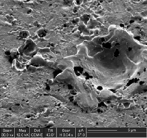
Titanium plasma spraying (coating)
2.1.2. AIR-ABRASIVE (BEAD-BLASTING) PROCESSING
One of the customary ways of the formation of the rough implant surface with considerable relief is air-abrasive processing. This method is based on the delivery of the hard ceramic particles moving at a great speed in the stream of the compressed air onto the implant surface. The surface roughness rate will depend mostly on the dispersibility of the particles used. It is well known that the contamination of the implant surface can interfere with osteo-integrative processes; that is why to avoid the contamination of the processed substrate it is necessary for the material used for granules production to be of chemically stable kind. As the materials for air-abrasive processing the aluminum oxide, titanium oxide and the calcium-phosphate ceramics particles are usually used.
The most popular material for bead-blasting processing is the aluminum oxide (Al2O3). It is possible to regulate the obtained surface properties with the help of particle-size analysis which makes it possible to select the aluminum-oxide ceramics particles with the necessary dispersibility rate. You should keep in mind that particles with high kinetic energy hitting the implant surface integrate into it and stay there regardless of the further ultrasonic cleaning, acid etching and sterilization. In some cases after the implant placement procedure these particles penetrate into surrounding tissues and influence the osteo-integrative processes. Even more than that, such kind of chemical heterogeneity of the implant surface can produce the negative effect onto the corrosion resistance of titanium in the tissues (Aparicio C et al., 2003).
As it was already mentioned, apart from the aluminum oxide the titanium oxide is also used for the air-abrasive processing. The titanium oxide particles with the average dispersibility of 25 microns are able to form the surface with the irregularities of 1-2 microns in size. Experimental studies where the implants that underwent machine processing were compared to the ones processed with the use of TiO² particles by means of bead-blasting method have shown that their placement results in the increase of the amount of possible bone-implant contacts observed at the microstructure level (Ivanoff C.J. et al., 2001). Other authors also share this point of view (Rasmusson L. et al., 2001) Higher viability rate observed for the ten-year functioning term of the implants processed by means of bead-blasting method is a great evidence of high clinical effectiveness of this method (Gotfredsen K. et al., 2001; Rasmusson L. et al., 2005).
Important clinical and X-ray criteria for the successful implantological treatment are the level of the marginal bone resorption around the implant. In the area around implants processed by means of bead-blasting method the rate of the parameter in question was significantly lower than in the control group, which can be regarded as the evidence of high effectiveness of this technology (van Steenberghe D. et. al., 2000; Astrand P. et al., 1999) Wennerberg et al. showed by his experiment carried out on rabbits approximately the same amount of the bone-implant contacts in the area of the implants processed by means of TiO2 and Al2O3. particles. In this case the mechanical stability of both kinds of implants was much higher than the one of the smooth implants produced by means of milling method (Wennerberg A. et al., 1999). This research showed that the screwing-in power of the processed implants increased in proportion to the roughness rate, at the same time when the amount of the newly formed bone tissue around implants wasn’t significantly changed. The research carried out by the above-listed authors corroborates the fact the increase of the implant surface roughness rate increases its mechanical fixation improvement but it still doesn’t influence the biological aspect of the implant stability (Abron A et al., 2001).
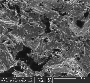
Air-abrasive (bead-blasting) processing(Dimitrovich D.A.)(1)
From the point of view of the optimization of conditions for the osteogenic cells fixation to the implant surface and subsequent osteogenesis it is well-known that the surface should be isotropic (that is have the same topology in all directions) and be characterized by the uniform porosity. This kind of surface can be formed with the help of air-abrasive method using the TiO2 particles of 60 -90 microns in diameter. (Mustafa K. et al., 2001). It is supposed that the surface with higher roughness rate formed by the use of particles of 300 microns in diameter (with Sa 1.4 parameter) doesn’t have any advantages in the context of bone substance formation, as in case of this approach the optimal roughness rates can be exceeded. According to one of the theories osteoblasts ranging between 10-12 microns in size are not able to identify the rather big micro-irregularities formed by means of this kind of processing, and they lose their capability for fixation and spreading and (as a consequence to this fact) their capability for differentiation.
The effective way of formation of significant implant surface micro-relief is the bead-blasting processing by means of the granules on the calcium-phosphate base, first of all such as hydroxyapatite and tricalcium phosphate. If we speak of the advantages of these substances, they are considered to be biocompatible, resorbable and have strongly pronounced osteoconductive properties. (Novaes A. et al., 2002; Piatelli M. et al., 2002; Mueller W.D. et al., 2003).
2.1.3. ACID PROCESSING OF THE IMPLANT SURFACE (ACID PICKLING)
As well as bead-blasting processing the pickling by means of highly concentrated acid solutions is applied to provide the considerable relief and roughness to the implant surface. The acids as HCl, H2SO4, HNO and HF are used for that. Acid pickling results in the formation of the microscopic pores of the size ranging from 0.5 to 2 microns in diameter (Massaro C. et al., 2002; Zinger O. et al., 2004). The increase of the osteo-integration rates as the result of acid pickling processing is the proven fact (Wong M. et al., 1995). Dipping of a titanium implant to the solution containing concentrated mixture of HCl and H2SO4 heated to the temperature of more than 1000С (being so called double acid pickling) results in the formation of pronounced implant surface porosity. The above mentioned processing method makes it possible to increase the osteo-integrative capacity of the implant surface, which has been proven by the data obtained for 3 years of its clinical studies (Cho S.A. et al., 2003). It was found that double acid pickling of an implant increases the osteo-conductive properties of the surface, in particular on the account of the increase of the osteogenic cells adhesion and fibrin adhesion rates in the course of the contact osteogenesis (Park J.Y. et al., 2000). In particular the process of formation of the coarse-fibered bone tissue (osteoid) with thin trabeculae on the implant surface is described here (Trisi P. et al., 2003). Some experimental studies demonstrate greater amount of bone-implant contacts and lower rates of marginal bone resorption in the implants processed by means of double acid pickling method if compared to the milled implants and implants processed by use of titanium plasma-spraying method (Davies J.E. et al., 1998; Trisi P. et al., 2002). Great attention is paid to the development of the acid pickling methods. The main effort is made in relation of providing the most favorable conditions for the fixation of the osteogenic cells to the implant surface, which can produce the positive effect on the de novo bone formation in the implant placement area (Trisi P. et al., 2002; Trisi P. et al., 2003). It is proven that the highest surface wetting ability rates are achieved by means of double acid pickling of the implant with the use of heated solution as in this way homogeneous surface with open porosity is formed (Cochran D.L. et al., 2002). High wetting ability rate of this kind of surface favors more intensive fibrin sedimentation, which in its turn produces the positive effect on the migration and adhesion of osteogenic cells (Novaes Jr A.B. et al., 2004; Papalexiou V. 2004). As it was already mentioned above one of the experimental studies carried out on mini-pigs shows that the amount of the bone-implant contacts (the factor being rather popular in a range of studies in terms of its informational content) was higher in the group where the implants with hydrophilic surface were used if compared to the group where ordinary implants processed by means of bead-blasting method or by means of acid pickling method were used.
The alternative chemical agents used for implant surface processing are fluoride solutions. Titanium easily interacts with fluoride producing soluble salt of the titanium fluoride (TiF4). There are some suggestions of the fact that dipping of the implants to the hydrogen fluoride (it is also possible to use hydrofluoric acid for this purpose) helps not only form the considerable micro-relief of the implant surface, but also results in the incorporation of fluorine ions into the implant structure which is, without any doubt, a positive factor for further osteo-integration (Ellingsen JE et al., 1995, 2004).It is shown that the osteoblasts differentiation on the surface of the implants being processed by means of this kind of chemical treatment is more intensive if compared to the control samples (Cooper L.F. et al., 2006). The resistance rate while pulling-out test of the implants processed with the use of hydrogen fluoride is also higher if compared to the implants used in the control group. In this way it is possible to assume the fact that the above-mentioned method of processing can impart some bio-active properties to the implant surface.
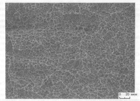
Acid processing of the implant surface (acid pickling)
As for the disadvantages of this method, they may include the negative effect of the chemical treatment produced on durability characteristics of the implants. The most dangerous phenomenon is such an undesirable side action as hydrogen acid brittleness or hydrogen embrittlement of metal, which results in a great amount of hair cracks on the implant surface, reducing the fatigue property rates of the endosteal implant part. Experimental data really proves the ability of titanium constructions, placed into the biological environment, to absorb the ions of hydrogen. The hydrogen embrittlement is mostly related to the formation of so called fragile hybrid phase, which results in the decrease of plastic characteristics of titanium. This phenomenon should be taken into consideration with the purpose of defining the reasons of the titanium implant fracture in the conditions of implant functioning in the oral cavity. (Yokoyama K. et al.,2002).
2.1.4. IMPLANTS PROCESSING BY MEANS OF ANODIZING METHOD (ANODIC OXIDATION METHOD)
The formation of the surface with micro- and nano-porous relief is possible to realize by means of potentiostatic or galvanostatic anodizing of titanium surface of the implant placed into the strong acids environment (H2SO4, H3PO4, HNO3, HF) with high conductance (200 A/m2) or voltage density. The result of anodizing is the formation of implant surface with the oxide layer of more than 1000 microns in depth. The presence of electrolyte (that is strong acids) results in the fact that under the effect of electric current there is dissolution of oxide layer in some implant surface areas and its thickening in the other ones. The dissolution of oxide layer takes place along so called convection lines and results in the formation of micro- and nano-porous titanium surface. (Sul Y.T. et al., 2001, 2005; Xiropaidis A.V. et al., 2005; Huang Y.H. et al., 2005). It is known that anodizing leads to the changes in surface microstructure and in the crystallinity rate of the surface oxide layer (Sul Y.T. et al., 2002). It is worth mentioning, that anodizing is a rather complicated process, and its effect depends on many parameters, and special attention should be paid to such ones as conductance, acids concentration, electrolyte temperature and electrolyte composition.
The result of the anodic oxidation of the titanium surface is the improvement of the biomechanical characteristics of bone-implant interactions, expressed in the significant strengthening of the implant fixated in the bone tissue, which is histologically proven (Sul Y.T. et al., 2002; Rocci A. et al., 2003). According to the data by Jungner et al. the viability of implants with the surface, processed by means of anodic oxidation method can be interpreted by the reliable mechanical linkage which is formed by the way of bone tissue growing into the pores of the implant surface and by means of biochemical bond of bone tissue to anodized titanium (Schupbach P. et al., 2005). Changes in the chemical composition of the implant surface after
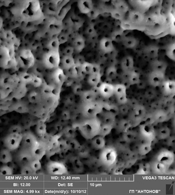
Implants processing by means of anodizing method (anodic oxidation method), Ñ…5000
anodizing consist in incorporation of the ions of magnesium, calcium, sodium and phosphorus (Sul Y.T. et al., 2002, 2004). There is the data proving the fact that the inclusion of magnesium ions into the oxide layer of the implant surface results in the increase of the implant resistance properties shown by the pulling-out test, if we compare it to the implants with the surface incorporated with other kinds of ions.
2.2 Application of the bioactive substances onto the implant surface for the optimization of the osteo-integration processes Inorganic substances
2.2.1 CALCIUM-PHOSPHATE CERAMICS COATING
There are following types of calcium phosphates: 1) Tricalcium phosphates (TCP); 2) Hydroxyl apatite; 3) Tetracalcium phosphate. Calcium-phosphate compounds possess pronounced osteogenic properties, provide the adhesion of proteins and bone cells, maintain ionic and covalent bonds with bone microphages. The biocompatibility rate of calcium-phosphate materials depends on its stoichiometry, crystallinity and porosity rates. In clinical practice two kinds of calcium-phosphate ceramics - hydroxyl apatite and tetracalcium phosphate - are used. The latter is more biodegradable than hydroxyl apatite.
After the introduction of the calcium-phosphate materials into the tissue the normal process of healing takes place. (de Groot K. et al., 1998). In this case the fibrous tissue is formed in a very small amount, and the material can come in direct contact with bone tissue.
Calcium-phosphate compounds strengthen greatly the bond with the bone tissue. The bond area is a thin (Daculsi G. et al., 2003) interface between the implant and the bone, containing highly mineralized substances and is practically free of collagen fibers. The disadvantage of this kind of osteo-integration is the fact that occlusal loading can result in the destruction of the implant bed because of the lack of depreciation. As for the advantages of the tricalcium phosphate ceramics, among them there are high biocompatibility, integration of the material with soft tissue and bone tissue, the minimal thermo- and electro-conductivity, as well as the modulus of elasticity close to the one of the bone tissue (30-120 GPa) and the limit slenderness ratio (15-120 MPa). It is also one of the materials which are described in a great number of research works and publications.
There is a great number of methods for the formation of the porous bio-active coatings on the implant surface (for example, the glue method, the isostatic pressing method, the knurling method, the method of synthesis, the electrochemical method, the investment casting method, the sputter coating method, plasma deposition method, etc.).
These factors initiate the healing of the bone tissue, favoring the macrophages activation, intensify the angiogenesis, and stimulate the formation of the collagen fleeces. It is worth mentioning that scientific publications abound in messages, stating the fact of the increased adhesion of protein substances to the surface of the implants, covered with calcium-phosphate materials. In vivo, bio-active coatings intensify the proliferation and the differentiation of the osteoblasts as well as the synthesis of extracellular matrix (Morris H.F. et al., 2000). The increased deposition of bone cells and growth factors on the calcium-phosphate coating surface are conditioned by their affinity with protein molecules of the implant site. This interaction is expressed through the complicated chemical processes: covalent and ionic bonds > electrostatic forces effect > hydrogen bonds, included into the hydrophilic groups, such as – ОÐ, – СООÐ, – NH2 > hydrophobic interactions (for example, adsorption of hydrophobic substances in water) > Van der Waals force.
The growth factors accumulated on the surface play one of the most important roles in the healing of the bone wound. These factors are constantly present in the native bone being at the same time the mediators of cell proliferation and differentiation, angiogenesis and mineralization, as in case of physiological conditions, as in case of reparative regeneration of the bone tissue. Being located in the damage zone they do not only attract osteogenic precursor cells, but also stimulate the synthesis of bone collagen proteins with osteoblasts as well as replenish their amount by means of their influence on the differentiation of their precursor cells that is osteo-progenitor cells.
Calcium-phosphate coating also has some weak points (Wheeler S. et al., 1996; Chang Y.L. et al., 1999). Among the most considerable ones there is the possibility of flaking of the coating from the titanium substrate in the course of implant functioning, regardless of reliable integration of material granules with the surrounding bone tissue. The nature of this undesirable effect is explained by the bond (not strong enough) between different phases of calcium phosphates on the implant surface (Lee J. et al., 2000). Hydroxy-apatite sprayed onto the implant surface by means of plasma spraying method is as a rule represented with the big crystallized particles so to say ‘walled-up’ into the highly soluble amorphous phase of the substance (Tinsley D. et al., 2001).
Flaking of the coating from the titanium support can take place immediately at the moment of the implant placement, especially in case of inconsistency in the size of implant and the size of implant bed, or in the conditions of high density of the bone tissue. It is rather problematic also to achieve satisfactory surface characteristics in case of thin implants or the implants having complicated form coating with calcium phosphates by means of plasma spraying processing method.
The new opportunities appear alongside with the development of so called biomimetic technology for the formation of the calcium-phosphate coating on the implant surface. With the development of the above-mentioned method it became possible to form the thin mineral layer of octacalcium phosphate - Ca8H2(PO4)6,5H2O, taking part in the formation of the bone tissue, as well as the layer of B-carbonated apatite having the structure and the architectonics very similar to the natural mineral phase of the bone tissue. Above-listed innorganic components are not present on the surface, formed by means of plasma spraying method. The definite know-how of this method is the fact that the formation of the surface is realized in two stages under the conditions very similar to the physiological ones. The essence of the technology is as follows. In the course of the preparatory stage after the sequential ultrasonic cleaning of the samples with the 70%- ethanol, acetone and demineralized water the 10-minute pickling of the surface with the mixture consisting of 2 ml of 40%- hydrofluoric acid (HF) and 4 ml of 66%- nitric acid (HNO3) in 1000 of water is carried out. Then the samples with rough surface are dipped into the so called ‘solution simulating the tissue fluid’ (SSTF), the composition of which is given in table 2.
Salts accurately weighed beforehand (±0.01 g) (reagent grade; Merck, Amsterdam) are dissolved in the demineralized water. The calcified solutions were buffered up to the physiological рР7.40 rate at room temperature with Tris-HCl 50 mM. After that the solution is filtered through the membrane0.22 micron thin (Millipore, Etten-Leur).
The second phase of samples immersion is carried out in polystyrene tubes, containing one of the SSTF (1-3). The tubes are sealed hermetically and then placed to the calibrated water-bath and are thermostated there at a temperature of 37◦ С. In 48 hours of immersion the samples are taken out, washed with demineralized water and are air-dried.
The research by Barrere F. et al., (1999) showed that the character of the coating at a great degree depends on the composition of the SSTF. The immersion of samples in SSTF1 results in deposition of the epitaxial crystals layer of octacalcium phosphate on the implant surface. The presence of ions of magnesium and carbonate in the SSTF composition produces the visible but multidirectional effect on the surface structure. The introduction of magnesium to the SSTF composition results in formation of the low-crystallinity calcium-deficient magnesium-containing layer of the apatite on the titanium surface. B-carbonated apatite precipitates on the implant surface by means of carbonate ions introduction to the octacalcium phosphate crystalline grid.
ORGANIC SUBSTANCES
2.2.2 Application of chitosan onto the implant surface
Chitosan (1-4.2-amino-2-deoxi-b-D-glucan) being a biopolymer, deacetylated derivative from polysaccharide of chitine is one of biologically active substances, attracting more and more interest for the last years. The prospective of this substance is its possible application in the domains of implantology, tissue engineering as well as for the purpose of development of special means for delivering of pharmaceutical composition to the body organs and tissues. This substance is found in the exoskeleton of some of the crustaceans, as well as in the insect cuticle, in the fungi cell wall and in the plankton. Chitosan has the polycationic hydrocarbon structure, similar to the hyaluronic acid and glucosaminoglycans; its degradation products (saccharides and glycosides) can be added to the glycoprotein or be released in the form of carbon dioxide (Suh F.J.K. et al., 2000). The biological effects of chitosan on the tissue include inhibition of fibroblasts proliferation, stimulation of the migration of polymorphonuclear and progenitor cells, activization of the nervous tissue and vascular tissue (Lahij A. et al., 2000). Besides, chitosan helps to form the bone and cartilage tissue, skin, and produces the antibacterial effect. All the above-listed properties make chitosan the prospective material for the application as the surface-active agent in the dental implantology domain.
|
Na (+) |
K(+) |
Mg (2+) |
Ca (2+) |
Cl (-) |
HPO4(2-) |
SO4 (2-) |
HCO3 (-) |
|
|
HP |
142.0 |
5.0 |
1.5 |
2.5 |
103.0 |
1.0 |
0.5 |
27.0 |
SSTF 0 |
142.0 |
5.0 |
1.5 |
2.5 |
147.8 |
1.0 |
0.5 |
4.2 |
SSTF 1 |
140.4 |
— |
— |
3.1 |
142.9 |
1.86 |
— |
— |
SSTF 2 |
140.4 |
— |
1 |
3.1 |
142.9 |
1.86 |
— |
— |
SSTF 3 |
140.4 |
— |
— |
3.1 |
142.9 |
1.86 |
— |
5 |
Table 2. Inorganic composition (mМ) of the human plasma (HP) and solutions simulating the tissue fluid (SSTF).
It is considered that the intensification of the cells adhesion on the surface of the implants coated with chitosan can be explained with the presence of the amino-groups of cationic nature in its substance composition. Prasitsilp et al. indicated to the higher rates of adhesion of mouse fibroblasts and hamster kidney cells to chitosan with the deacetylation rate of 86-90% with the high content of D-glucosamine if compared to the chitosan with the deacetylation rate of 76 – 81% (Prasitslip M. et al., 2000). Chatelet et al. found out that the level of keratinocytes and fibroblasts adhesion to the chitosan layer with the deacetylation rate of more than 85% was higher than in case of chitosan layer with lower deacetylation rate (Chellat et al., 2000). An interesting fact is that fibroblasts were fixing better than keratinocytes. But keratinocytes surpassed the fibroblasts in relation of their proliferative activity on chitosan layers with high deacetylation rate. The authors assumed the fact that too intensive adhesion of fibroblasts contributes to the inhibition of their proliferation. Above-mentioned data shows that the increase of the deacetylation rate leads to the increase of positive charge density of chitosan, increasing its capability for attraction of negatively charged cells. This fact can play an important role in the formation of tissue response in case of wound healing process, as it is well-known that introduction of chitosan with high deacetylation rate to the wound results in any case in the formation of the connective scar.
The interaction between the positively charged chitosan polymer and negatively charged components of red blood cells and platelets, responsible for coagulation and hemagglutination in the process of blood clot formation was discovered (Rao et al., 1995). Then the researchers demonstrated the fact that as a result of chemical and radiation sterilization as well as the result of autoclaving the decrease of chitosan wetting ability rate takes place, which in its turn reduces the level of red blood cells hemolysis. Unfortunately these researchers didn’t provide any information on the deacetylation rate of the chitosan they used in their studies.
The suggestion about the fact that chitosan intensifies the osteointegrative processes is proved by Bumgardner et al. (2003), as they showed that the

Calcium-phosphate ceramics coating.
chitosan coating of implants contributes to the strengthening of the human palatinal embryonic mesenchymal cells attachment, increases the degree of adsorption of albumin and fibronectin on their surface unlike the cases when the implants without chitosan coating are used.
2.2.3 APPLICATION OF THE MORPHOGENETIC PROTEINS ONTO THE IMPLANT SURFACE
Starting the reaction cascade, signaling molecules (being the bone morphogenetic proteins or BMP) related to the super-family of transforming growth factors β, play one of the most important roles in osteo-integration process, starting with the primary tissue response and finishing with the termination of the secondary change. As a rule, growth factors are subdivided into groups according to their function depending on the fact what reparation component they influence (the cell differentiation or the cell proliferation). The influence of above-mentioned polypeptide factors is dose-dependent and changes according to the concentration of this or that factor in the tissues.
These multifunctional molecules are able to cause the ectopic bone formation and modulate the expression of some proteins with osteoblasts. Going into detail on the influence of BMP on the osteo-integration processes we should mention that these factors contribute to the intensification of the expression on the implant surface of the osteoblastic line cells of specific receptors, favoring the adhesion of the cells to the implant surface. For example, BMP-2, well-known for is functions, produces the intensifying effect on the cytoskeleton of osteoblastic cells, on the organization of extracellular matrix and increases the rate of cells fixation to the titanium surface. Besides, there are research works showing the possibility of BMP-2 integration into the composition of bioactive coatings applied onto the titanium surface, an when the substance still stayed active.
Although these substances are called bone proteins their activity is not restricted with their influence on the bone tissue. BMP-1 doesn’t take part in the growth and differentiation of the osteogenic cells, it is the protease which splits collagen fiber and it is referred to the separate group. Activity being not typical is carried out by BMP-3 (osteogenin). Unlike all the other proteins of the BMP family this polypeptide acted as the bone formation inhibitor in a series of experiments, being the antagonist for other BMP (Aaron Daluiski et. al., 2001). BMP-7 famous also as osteogenetic protein-1 was initially identified as cartilage and bone tissue formation regulator. Later it was found out that this protein stimulates the growth, the chemotaxis, the differentiation and the apoptosis of different kinds of tissue, here including epithelial, mesenchymal, hematopoietic and nervous ones. Besides, this mediator is considered to produce the protective effect on the renal tissue and take an active part in the morphogenesis of this body organ. (Zeisberg M., 2006).
The specific feature of the bone morphogenetic proteins (if we compare them to the other growth factors) is their ability to transform the cells of the connective tissue into the osteo-progenitor ones, which was not discovered in the course of studies of the insulin-like, fibroblast, epidermal and other growth factors. In this way these substances not only produce the stimulant effect on the mitotic activity of cells, favoring the cell division, but also influence the tissue morphogenesis. It is realized by means of binding of bone morphogenetic proteins to the specific receptors which can be of two kinds – BMP receptors of type 1 and BMP receptors of type 2, being located on the osteo-progenitor cells surface. In our days minimum three variants of receptors of type 2, able to interact with osteo-inductive BMP molecules, including BMP-2, BMP-4 and BMP -7 have been identified.
At the market of the osteoplastic materials the preparation of new generation containing bone morphogenetic protein in its composition was tested quite recently. The Infuse (Medtronic Sofamor Danek) preparation is produced in the form of collagen sponge carrying the recombinant BMP-2, synthesized by the ovary cells of Djungarian hamster. The ovary cells of Djungarian hamster is the time-tested and safe model functioning as a kind of a factory for the synthesis of various proteins. The recombinant BMP-2 obtained as a result of gene engineering development is purified by ultra-filtration method and then undergoes freeze drying. As for the practical work, the preparation is provided in the form of white crystalline powder which should be wetted for 5 munites before use with the sterile water. Then it is applied onto the carrier to which it should be fixated for 15 minutes. At the present time it if permitted to use this preparation in the domain of general orthopedics, especially for treatment of the degenerative changes of intervertebral discs. At the present time the tests are held in relation to the usage of this material for dental implantology needs, notably for the SFE procedure.
There are a lot of research works related to the formation of bioactive coatings with pronounced osteo-inductive properties. One of the most often studied agents in this domain is the BMP-2. The practical attraction of this substance is related to the certain technological difficulties in the course of their application. The actual problem is the choice of the way for BMP-2 application onto the implant surface. The above-mentioned protein molecules have low affinity rate in relation to the metal alloys and in case of direct impregnation of the implant surface with the bioactive substance containing BMP in its composition they are dissolved very fast in the tissue liquid and then inactivated with specific proteins (noggins). This process can take from several hours to several days and it doesn’t give the possibility of the full realization of therapeutic potential of this substance. To eliminate this effect some researchers made an attempt to increase the concentration of the BMP placed on the implant surface to up to non-physiological limits, which didn’t have the desirable effect on the reason of retention of the increased speed of protein molecules release into the tissues. This rapid release of big amounts of the above-mentioned water-soluble substance in the tissue leads to the active binding of protein molecules by the bone collagen and other proteins of the extracellular matrix, minimizing the effect produced by BMP on the osteogenesis processes. Another disadvantage is the fact that apart from some intensification of the osteogenesis in the dental implantation area BMP containing in high concentrations in the tissues contributes to the attraction of osteoclasts and intensification of the osteoclasts activity, increasing considerably the resorbtion of bone tissue and producing the catalytic effect on the implant coating if there is any. BMP show higher affinity (if compared to the metal surface) to the calcium-phosphate materials that can play the role of three-dimensional matrix for the transfer of biologically active substance from the implant surface to the tissues. In this case the biologically active substance release to the environment happens gradually and at a lower speed. Thanks to the development of the technology of biometrical coating formation which was already mentioned in the previous section, it became possible not just apply but introduce BMP into the structure of the calcium-phosphate crystalline grid on the implant surface, making it possible to achieve even more metered release of bioactive substance into the tissues. The experiment shows that this process takes place synchronously with the dissolution of the calcium-phosphate matrix on the implant surface. It means that in 5 weeks with degradation of the 60% amorphous calcium phosphate applied on the surface of the titanium disks implanted under the skin of laboratory animals the level of BMP-2 on the implant surface stays the same. The diffusion in the tissue of the most part of this protein substance is directly conditioned by the osteoclastic resorption of the mineral matrix on the implant surface. This process has the common nature with cell-tissue interactions in the period of bone tissue remodeling, when the gradation of the mineral matrix takes place simultaneously with the release of the growth factors from it.
In the course of the experiment on white Japanese hares Chinese scientists carried out the comparative analysis of the osteo-integration rates of the usual implants and the implants, impregnated for 30 minutes in 2 ml of sterile water, containing 1.0 mg of the BMP-2 recombinant (Lan J. et al., 2007). It was found out that in 12 weeks after the implant placement in the course of pull-out test more effort was made for pulling out the implants with modified surface if compared to the non-impregnated implants 36.5 ± 2.02N to 27.63 ± 1.31N correspondingly. It proves that more reliable implant insertion can be provided by the implants with the surface impregnated with BMP-2 recombinant. The histomorphometric studies comprising the count of labeled bone sections, belonging to the implant also showed higher rates in the main group (15%) compared to the control group (11%), which is the evidence of more rapid maturation and mineralization of the bone tissue around implants, impregnated with bioactive substance.
Using the histomorphometric method, the same group of scientists carried out the comparison of the surfaces impregnated with different bioactive substances. 64 titanium implants of a standard size covered with a polylactide acid layer were implanted into the femoral bone of 16 adult Japanese hares. All the implants were subdivided into 4 equal groups. The 1st group consisted of the implants wetted with 2ml of sterile water containing 1.0 mg of recombinant BMP-2 and 200 mg of recombinant fibroblast growth factor (bFGF). The implants of the 2nd group were wetted with the same composition but unlike composition used for the 1st group the liquid used in the 2nd group contained 250 mg of recombinant human insulin-like growth factor-I (IGF-I) instead of bFGF. In the 3rd group the implants wetted with 2 ml of sterile water containing recombinant BMP-2 were used. The 4th group was a control one and the implants used for it were impregnated into the pure solution of sterile water without any bioactive components. The impregnation of all the implants in the solution took 30 minutes, and after that the samples lyophilization (freeze-drying) and preservation at the temperature of -20◦С were carried out. To avaluate the osteogenesis rates during the implant survival term the fluorescent bone markers were implanted to animals. In 4 months after the implantation the intravenous infusion of the calcein green in the dosage of 20 mg of calcein for 1 kg of weight of an animal was carried out. In 8 weeks after the implantation the intravenous injection of 20 mg of alizarin red for 1 kg of weight of an animal was carried out. In 12 weeks after the start of the experiment animals were withdrawn from the experimental groups and the quantitative analysis of the osteogenesis rates in the implant area was made. The implants were removed together with the surrounding tissues and were immediately fixated for 7 days in the 10% solution of buffered formalin at a temperature of 4˚С. The samples were dehydrated with alcohol with the concentration increasing from 70% to 100% and then were dipped into the methyl methacrylate plastic. After the polymerization the cutting of the samples into thin sections 50 microns thick along the long axes of the implant was carried out with the help of diamond disc. The samples studies were made with the help of confocal laser scanning microscope.
As a result of calculations the statistically significant difference between the samples from different groups was found. The new formation of bone tissue in the second group (BMP-2 + IGF-I) was more remarkable than in the 3rd group (BMP-2) but this statistically significant difference was observed only in 8 weeks after the implant placement. There was no any remarkable difference between the 1st and the 2nd group rates although in 4 weeks of experiment the insignificantly smaller amount of the newly formed bone tissue was observed in the 2nd group. The 4th (that is the control) group demonstrated the worst (the lowest) rates.
It is difficult to overestimate the importance of the growth factors and bone morphogenetic proteins in particular. But it is still worth mentioning that the clinical application has several restrictions. The BMP molecules sent very subtle and short signals to their targets, initiating the cascade of cell-tissue reactions. These effects vary significantly depending on the cells type, their location and environment, their differentiation rate and (what is especially important) the presence of other morphogenetic proteins in the area of their activity. That is exactly why the fact that every factor taken individually doesn’t produce the effect of significant intensification of the implants integration in the bone tissue is not surprising.
2.2.4. APPLICATION OF THE ADHESIVE MOLECULES ONTO THE DENTAL IMPLANTS
A great number of scientific research works is devoted to the studies of the cell adhesion effect on the implant surfaces and on their differentiation and further proliferation. The possible way for the osteoblastic cell adhesion potentiation is the immobilization of peptides, containing arginine-glycine- aspartic acid, on the implant surface (Rezania A., Healy K.E., 1999). The recent studies showed that tetra-cell molecules of adhesion, being the adhesive substances, contribute to the differentiation of the osteoblast-like cells, cultivated on the non-organic bone mineral matrix of the bovine nature. The tetra-cell adhesion molecule is the recombinant protein, containing arginine-glycine-aspartic sequence in the tenth type of the III domain, proline-histidine-serine-arginine-aspartic sequence in the ninth type of the III domain of fibronectin, tyrosine-histidine sequence and glutamic acid-proline-aspartic acid-isoleucine- methionine sequence fas-1 domain βig-h3. Arginine-glycine-aspartic sequence is able to bind to some of the integrins on the cell surface; one of the most important of them is α5β1. This interaction shows the stimulating effect of the above-stated sequence on the adhesion of the osteoblast-like cells. (M. Kantlehner et.al., 2000). The proline-histidine-serine-arginine-aspartic sequence in the ninth type of the III domain produces the synergistic effect in context of the intensification of the adhesive affinity of the arginine-glycine-aspartic sequence. There is the data relating to the fact that both of these sequences of the tetra-cell molecule are identified by the integrin α5β1 on the surface of the osteoblast-like cells (Aota S. et al., 1994). βig-h3 is the protein of the cell adhesion; it comprises 4 internal repetitive domains being called fas-1. The expression of this domain in some types of cells at a greater degree is induced by the transforming growth factor β. The recent studies showed that the application of e inorganic bone mineral matrix of the of the bovine nature in combination with substrate on the base of tetra-cell molecule adhesion produces the significant stimulating effect on the formation of the mineralized bone substance in case of replacement of the defects of the hare skull bone tissue (J.H. Lee et al., 2006).
The scientists from Daegu Institute (South Korea) studied the biological effects of tetra-cell adhesion molecules application on the micro-porous surface of dental implants. As the phenomena being the subject of studies the effect of the above-mentioned substance on the re-implantation osteogenesis in the conditions of the compromised state of the bone bed. The implants were placed into the femoral metaphysic, which is characterized by a small amount of spongy substance. The implants were placed at a minimal level of primary stability which was achieved by means of formation of the canal with the insignificantly larger diameter if compared to the implant diameter. In the control group the implants made of commercially pure titanium (Grade 2) processed by means of air-abrasive method with the use of the hydroxyl-apatite particle granules of 100 microns in size were used. Then the surface passivation with nitric acid according to the specification ASTM (American Society for Testing and materials) was carried out. After the above-mentioned processing the implants used in the main group were placed into the phosphate-buffered medium. Immersion was carried out for 24 hours at room temperature at 100 mg of tetra-cell adhesion molecule solution for one square mm of the implant surface. In the course of previous studies this concentration of tetra-cell adhesion molecules showed maximum effectiveness in relation of the developmental potential of the osteoblast-like cells. For the removal of non-bound molecules each of the implants was washed three times with phosphate-buffered solution and then dried in the chamber by means of cross-border (laminar) airflow for 24 hours. The implant surface roughness rates of the main and the control groups didn’t have much difference, which is proven by the profilometry data. All the implants were sterilized by means of gamma-irradiation.
The experiment was carried out on 7 adult New Zealand white rabbits. The implants were placed into the femoral bone of a rabbit (one implant to each of femoral bones). The excess of the prepared implant bed size in relation to the implant apical diameter was 0.15 mm. The gap width was smaller near the apical part of the implant which made it possible to achieve the minimal stability rate of the implant. This interrelation was achieved by means of specially created bone-drilling burs. The wound was tightly sutured.
The histomorphometric study showed that the bone tissue healing process had no special features and was not accompanied with any inflammatory reactions. In 8 weeks of healing the implants as of the main as of the control groups had contacts with the cortical bone in the area of the upper implant part; the new formation of the bone tissue in the area of the bone-marrow area was also studied.
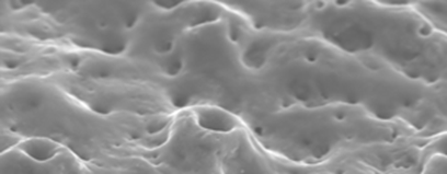
Application of chitosan onto the implant surface
2.3 The ways of modification of the surface, suggested by the leading manufacturers of the dental implants
2.3.1 THE SLA AND SLACTIVE COATINGS BY THE STRAUMANN COMPANY
The SLA surface is formed by means of air-abrasive surface processing with the use of coarse-grained corundum particles of 0.25 to 0.5 mm in size. It results in the formation of macro-rough relief of the implant surface. After the post-process machining the implant is dipped for several minutes into the container with high-concentration acids mixture (HCl/H2SO4), increasing the temperature alongside with it. As a result the micro-hollows and craters of 2-4 microns in diameter are formed on the implant surface; they expose onto the micro-irregularities, formed at the stage of air-abrasive processing. In this way the surface doesn’t become micro-porous and the closed cavities which make the surface less susceptible to the bacterial invasion are not formed.
By means of X-ray photoelectron spectroscopy it was found that the obtained SLA coating is the pure titanium oxide (TIO2). This method is
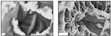
The SLA and SLActive coatings by the Straumann Company
able to track down the first several atomic layers of the surface chemical composition that is the layers that are directly involved into the interaction with tissue liquid and cells.
The SLActive surface is a modified variant of the SLA surface. The wetting ability rates and the chemical activity rates of this surface are achieved by means of implant processing in the presence of the nitrogen (N2) gas environment and then implant dipping into the container with isotonic sodium-chloride solution. There is the data showing that the presence of isotonic solution of NaCl on the titanium surface produces the protective effect in relation of the carbonates and organic impurities, containing in the atmosphere. In this case the surface stays clear and retains its activity rate (Buser et al., 2004). X-ray photoelectron spectroscopy demonstrates the lower level of the contamination of the modified surface with foreign substances. The content of the carbonate on the SLA surface corresponds to the level of the contamination of this substance on the other commercially available kinds of surfaces, coming into contact with atmospheric oxygen for a certain period of time. The high wetting ability rate of the SLActive surface is proven by means of contact angle determination method.
2.3.2 THE TIOBLAST AND OSSEOSPEED SURFACES BY ASTRA TECH COMPANY
The TiOblast coating is created by means of bead-blasting treatment of the implant surface with dioxide titanium particles. This method of surface processing was developed in 1990 and became the predecessor of the method of the OsseoSpeed surface formation which appeared later. As a result of bead-blasting treatment (Grade 4) of the implant with dioxide-titanium (TiO2) particles of 25 microns in size, the moderately rough surface with the Sa parameter of 1.10 microns is formed (Wennerberg, Albrektsson, 2000). This way of surface modification results in the formation of the topology with high relief uniformity rate. The great effectiveness of this method is proven by the studies demonstrating the higher level of bone-implant contacts and higher degree of bone tissue regeneration in the defects area, if compared to the implants that underwent machine processing. The implants with TiOblast surface are also famous for their high mechanical mechanical stability in the bone tissue, which is proven by the resonance frequency analysis.
The latest TiOblast technology development is the changing of the physic-chemical properties of the implant surface by means of the method of fluorination with diluted hydrofluoric acid (Ellingsen, 2004). One of the research works shows that modification of implants by means of the fluorination method increases significantly the conic implants fixation rates in rabbits in 4 and 8 weeks of the implant survival. (Ellingsen,1995) The studies of the osteo-integration rates carried out by means of light microscopy showed that the surface modified by the use of fluorine demonstrates tighter junctions with bone tissue, if compared to the non-modified surface. In the course of in-vitro studies it was shown that fluorine-modified titanium surface adsorbs the calcium phosphate (CaP) crystals from the solutions saturated with these substances. This phenomenon was not observed on the non-fluorinated implants surface.
It is well known that the fluoride compounds are able to stimulate the new formation of the bone tissue, before everything by means of the intensification of osteoblasts proliferation (Bellows C.G. et al., 1990). It is supposed that this effect is based on their ability to release fluorine from the implant surface as a result of phosphate-exchange reactions at the early stage of the wound process at the boarder of the implant and the bone tissue. In this way, the implant surface, due to its affinity to the calcium phosphate compounds plays the role of the object for precipitation of these substances intensifying the bone-implant interactions and as a result increases the stability of the implant itself.
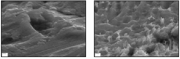
The TiOblast and OsseoSpeed surfaces by Astra Tech Company
2.3.3 TIUNITE SURFACE BY NOBEL BIOCARE COMPANY
The anaphase form of the titanium dioxide (TiO2) is one of the well-known crystalline forms of TiO2. As a rule this form of titanium dioxide is created by means of the method of thermal oxidation of titanium or by means of anodizing. This form of titanium dioxide demonstrates photocatalytic activity in case of A ultraviolet irradiation, which is a basis of its bactericidal properties and its ability for decontamination; it also increases dramatically its wetting ability rate.
TiUnite surface is created by means of anodizing method that is electrochemical process which results in the increase of the depth and roughness of the oxide layer on the implant surface. An implant is dipped into the electrolyte and it becomes the anode in the electrochemical cell. When the potential is applied to the sample, the ion transport of charge is passed through the electrochemical cell and the electrolytic reaction takes place on the anode resulting in the increase of the oxide layer depth. This technology is applied for the production of the Branemark System implants
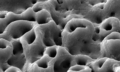
TiUnite surface by Nobel Biocare Company
(Nobel Biocare USA, Yorba Linda, CA) being the implants made of the commercially pure titanium. The roughness rate of the implants processed according to the TiUnite technology increases from 1 to 2 microns (the distance from the unevenness top to its bottom) in the coronary part of the implant and from 7 to 10 microns in its apical part. The name of the electrolyte used in the above-mentioned technology is not reported by the manufacturer company.
2.3.4. THE OSSEOTITE AND NANO TITE SURFACE BY 3I COMPANY
These methods of implant processing are developed by the Implant Innovations, Inc. Company (Palm Beach Gardens, FL). The Osseotite technology consists in double acid pickling of the rough implant surface by the use of acids solution (15% HF) and then with the mixture of acids H2SO4/HCL in the 6:1 ratio at the temperature rising from 60˚ to 80˚С for 3-10 minutes.
The comparative multicentre meta-analysis of the rate of survival of the implants that underwent the machine processing and the implants processed according to the Osseotite technology in the conditions of low qualitative characteristics of bone tissue showed the advantages of the implants with modified Osseotite surface. The data provided here are based on 8 prospective studies, analyzing the statistics of the viability of the 2614 Biomet 3i implants that underwent the machine processing and were implanted in 22 private and University clinics, and 2288 implants with Osseotite surface, that were implanted in 25 medical centers. Then there was 4-year follow-up period. As a result it was found that in all the tissue types the general survival rate of the implants that underwent the machine processing is 92,7% for 4 years. By the way, implants of this kind placed into the bone tissue of medium or high density showed the cumulative survival rate of 93.6% if compared to the rate of 88,2% for the implants that were placed into spongy bone tissue. The above-mentioned analysis shows that the effectiveness of the Osseotite implants application doesn’t depend on the qualitative characteristics of the bone tissue and in general these implants demonstrate the higher viability rates than the implants that underwent machine processing.
Disclaimer from Guident
The views and opinions expressed in articles published in Guident are solely those of the authors and do not necessarily reflect the views of the editors, the editorial board or the publisher, Ivory India. Guident does not endorse any product, technique or treatment described. Clinical content is intended for qualified dental and medical professionals and is not a substitute for individual clinical judgement. Guident, its editors and the publisher accept no liability for any consequence arising from the use of information published in the journal.
How to cite this article
Dr. Vologin G. A MSMSU. Methods of the Dental Implant Surface Modification. Guident. 2014 Jul;7(8). Available from: https://guident.in/articles/implantology/methods-of-the-dental-implant-surface-modification.html