"Implant Overdenture- a Viable and Cost Effective Prosthodontic Treatment Consideration"
Department of Prosthodontics, Seema Dental College and Hospital, Rishikesh, Uttrakhand
ABSTRACT
Edentulism is a problem that affects millions of people worldwide. A wide variety of new treatment modalities for restoration of edentulism with implants have given boon to clinicians. Implant supported overdenture has been used to overcome the inherent problems of mandibular conventional complete denture like retention and stability. Implant has become treatment of choice in both completely and partially edentulous patients. This article describes a simple, economical method of conversion from conventional complete mandibular denture into implant retained overdenture so as to improve the clinical outcome that is best suited for the patient.
KEY WORDS :
Conventional complete denture, Residual ridge resorption, Implant supported overdenture
INTRODUCTION
:
Conventional complete dentures rely upon the condition of residual alveolar ridge and mucosa for the purpose of retention, stability and support. Due to continued bone resorption, the adaptation problems i.e. loosening of dentures are observed with a higher incidence for mandibular rather than the maxillary dentures.1 The use of implants has a great impact on the prosthodontic treatment of the edentulous condition. Mandibular overdenture supported by only a few intraforaminal implants are regarded nowadays as a better treatment modality for geriatric patients.2 The indication comprises a large segment of older patients who loses their teeth in advanced age and also the patients who had complete denture for many years and lose their motor skills and no longer able to wear complete dentures. The problem is observed much more for the edentulous mandible than the maxilla.
The number of implants to be placed depends on the type of prosthesis and the choice of prosthetic design. Conversely, the number of implants that can be placed with respect to anatomic-morphologic conditions will also determine to a certain degree, the type and design of prosthesis. Misch3 proposed 5 prosthetic treatment options available in implant dentistry that are FP 1, FP 2, FP 3, RP 4, RP 5. FP 1 is a fixed prosthesis that replaces only the crown and looks like a natural tooth. FP 2 is a fixed prosthesis that replaces the crown and a portion of the root, crown contour appears normal in the occlusal half and elongated or hyper-contoured in the gingival half. FP 3 is a fixed prosthesis, replacing missing crowns and gingival colour and a portion of the edentulous site, prosthesis must often use denture teeth and acrylic gingiva, but may be porcelain to metal. RP4 is the removable prosthesis that is supported completely by implants and this design requires about 5 to 6 implants in mandible and 6 to 8 implant in maxilla. RP 5 is a removable prosthesis that is supported by both implants and tissue.
Additionally, the size, curvature and shape of the ridges determine the distribution of the implants over the arch. Economic factors may dictate the use of an overdenture supported by a few implants, notwithstanding possible additional maintenance costs associated with a removable design. Currently, controversy exists concerning the relative merits of fixed detachable prostheses versus overdenture prostheses. This is particularly evident with regard to maintenance, complications, and longevity.4 -6 Hemmings et al7 found that overdenture prostheses presented fewer maintenance requirements and complications than fixed prostheses over a study of 5 years. In contrast to maxillary overdentures, treatment outcomes with mandibular overdentures appeared to be more successful and are better documented, particularly for elderly patients.8-10 The aim of this article is to present a simplified approach to resolve the problems associated with conventional mandibular denture due to a lack of comfort, retention, stability, compromised esthetics and inability to chew.
CASE REPORT
A 60-year-old male patient reported to the Department of Prosthodontics at the Seema Dental College and Hospital with a chief complaint of difficulty in chewing with the previous denture. Dental history revealed that patient had undergone uneventful full mouth extractions 1 years back due to periodontal involvement and had made two pairs of conventional dentures, but was not satisfied functionally and psychologically. There was no history of systemic illness.
Intraoral examination revealed Misch Type 2 division of edentulous arch with U-shaped hard palate. Mandibular ridge was irregular with sufficient ridge height and width between the mental foramen and also with normal labial, buccal mucosa and floor of the mouth. No hyperplastic or flabby tissue was found. Class I ridge relationship with inter-ridge space of 12mm was observed. (Fig 1)
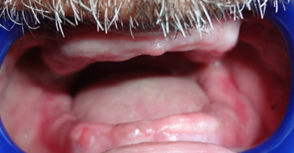
FIGURE 1: Pre-operative view
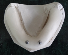
FIGURE: 2 Metal spheres of 5mm diameter incorporated in templates
All vital signs and blood investigations were within the normal range. For radiographic investigations Orthopantomogram (OPG )was done and to calculate the magnification error a template was fabricated with incorporation of metal sphere of diameter 5mm and thus effectively evaluated the distortion of image by using simple equation (A real/ A-PR=D-real/D-PR). (Fig 2, 3)
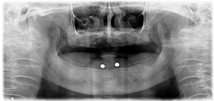
FIGURE: 3 OPG showing metal sphere to check the magnification errors
PROCEDURE:
- After completion of examination and investigations. Complete denture for maxillary and mandibular arch was fabricated in conventional manner. Post insertion issues were addressed favourably and patient was recalled after 15 days for implant surgery. (Fig 4)
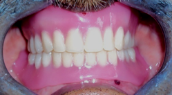
FIGURE: 4 Conventional complete dentures in place
2.Soft and hard tissue height was re-evaluated clinically and compared with radiographic diagnostic image to visualize a possible anterior loop of mandibular neurovascular canal. The goal was to place implant at least 2mm in front of mandibular nerve.
3.Bilateral mandibular block was administered to the patient (Lidocaine 2% with 1:200,000 Adrenaline).
4.OPG and Bone gauge were used to standardize length and width of implant (Adin Dental Implant System) that was 13.5mm and 3.35mm respectively.
5.Crestal incision was made with B.P handle with blade no. 15 and flap was reflected with Periosteal elevator. At required sites, Rongeur forceps was used to remove sharp edges, irregularities and no. #6 round bur was used to create dimple in the recipient site so as to allow easy initiation of twist drill hole. (Fig 5)
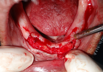
FIGURE: 5 Crestal Incision
6.Watson RM, Davis DM. Follow up and maintenance of implant supported prostheses: a comparison of 20 complete mandibular overdentures and 20 complete mandibular fixed cantilever prostheses. Br Dent J 1996;181:321-7.
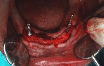
FIGURE: 6 Guide pin placed in prepared hole and angulation was checked to aid in parallel implant site preparation
7.After checking parallelism, the depth drills of diameter Ø 2.8mm, Ø 3.2mm and Ø 3.65mm were used.
8.Two implants were placed in the mandible between the anterior interforaminal regions i.e. one each on predetermined site. Flap was closed after placing healing abutment with interrupted sutures. (Fig 7, 8)
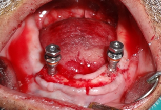
FIGURE: 7 Implant placed in the prepared site with torque wrench
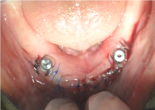
FIGURE: 8 Closer of flap after placing healing abutment with interrupted sutures.
9.Mericske-Stern R, Steinlin ST, Marti P, Geering AH. Peri-implant mucosal aspects of ITI implants supporting overdentures. A five-year longitudinal study. Clin Oral Implants Res. 1994; 5: 9-18.
10.Cune MS, de Putter C, Hoogstraten J. Treatment outcome with implant retained overdentures. Part 2: patient satisfaction and predictability of subjective treatment outcome. J Prosthet Dent. 1994; 72: 152-8.

FIGURE: 9 Post-operatives view after 3 month
11.Healing abutments were replaced by the ball abutment into the implant with Torque Wrench or Ratchet Wrench (recommended torque is 25 N/cm). Metal housing with nylon ring was placed over the ball abutment and block-out was done using sticky wax and rubber sheet. (Fig 11)

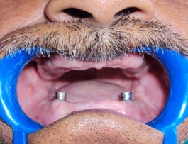
FIGURE: 10 Replacement of healing abutments by the ball abutment & metal housing with O-Ring

FIGURE: 11 Blocking Out
12.Pressure-indicating paste was used to transfer the marking from the metal housing to the tissue surface of the mandibular denture prosthesis and marked area was relief in denture to accommodate passive fit when seated over the attachments. (Fig 12)
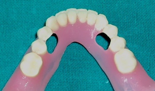
FIGURE: 12 Marked area of metal housing was relief to accommodate passive fit when seated over the attachments.
13.Self cure acrylic resin was mixed in conventional manner and injected in to the relief area and denture was seated into place.
14.Processed implant supported overdenture (RP-5) was removed, finished and polished once self cure acrylic has set. (Fig 13).
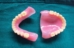
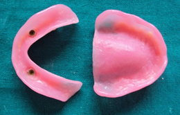
FIGURE: 13 Implant supported mandibular overdenture showing o-ring with metal housing.
15.Implant supported mandibular overdenture opposing the maxillary conventional denture was inserted in patient mouth. (Fig14). Oral hygiene and maintenance instructions were given. There was increased patient acceptance. Patient was recalled and necessary postinsertion adjustments were done for a regular period of time. There was an improvement in retention, stability, function, and also high degree of satisfaction was observed with mandibular implant supported overdenture in place. Esthetics was restored and there was also rebuilding of self confidence in patient.

FIGURE 14: Post operative view
DISCUSSION
The goal of modern dentistry is to achieve integration of dental implants and prosthetic treatment which replaces a missing tooth or teeth and lost supporting structures, as well as to restore proper esthetics and function. In order to achieve this goal, it is necessary for the patient and dentist to be able to visualize the outcome of the final prosthetic results, prior to surgical treatment. Therefore it is essential for the dentist to have a thorough understanding of all phases of treatment in order to diagnose, treatment plan and restore natural appearance.
The use of implants has a great impact on the prosthodontic treatment of the edentulous patient. A variety of prosthetic designs associated with implant prostheses can be observed, and some new designs have emerged in response to the specific clinical conditions of the edentulous jaw provided with implants. Valid clinical methods and treatment strategies – involving the implant technology exists to ensure the quality of prosthetic reconstruction. Hence, with regard to some special implant indications, specific prosthodontic principles are needed.
In many cases the treatment of the edentulous maxilla will require more elective procedures than are necessary for the mandible, particularly with respect to the following criteria: degree of atrophy of the residual jaw; prospective location of the implants and inclination of the implant axis; tissue volume dimensions; facial morphology; esthetics, function and phonetics. In general, the following criteria will determine the treatment planning of the edentulous jaw: The prosthetic design will depend on the distribution of the implants over the arch, their location and their number; The natural dentition or type of prosthesis in the opposing jaw will influence the implant-prosthodontic design and the intermaxillary relationship has to be considered; The occlusal scheme is influenced by all these factors; Esthetic considerations have to be involved, In the context of these criteria, implant prostheses must be planned, designed and managed for the edentulous jaw.
Batenburg et al11 evaluated 60 mandibular overdenture patients divided into two groups: first group was treated with 2 implants and other with 4 implants and found that no significance difference in peri implant health. Even with increasing the number of implants from 2 to 4 there is only a slight improvement in retention, stability and function as reported in retrospective study done by Mericske-stern’s 8 In 1987, Van Steenberghe et al proposed placement of two implants in edentulous mandible with success rate of 98%.12 Implant supported mandibular overdentures using two isolated abutments are successful treatment options but they can be demanding in periodic continuing care.13, 14 The Photoanalysis done by Kenney and Richards indicated that ball/O ring transfers less stress to the implants and also minimized the denture movement.15,16 It is mostly used as it is economic, less technique sensitive and easy to clean when compared to bar attachment.17 A variety of techniques have been reported in the literature for the incorporation of attachments in implant supported overdenture with unsplinted or individual abutments. Bulent and volkan described a 2- stage impression technique that records mucosa in functional state and placement of implant components accurately but disadvantage of this technique is that it is time consuming.18
The primary reason to consider dental implant for the replacement of the missing is the maintenance of the alveolar bone. The dental implant placed into bone serves not only as an anchor for prosthodontic device, but also as one of the better preventive maintenance procedure. An endosteal implant can maintain bone width and height as long as implant remains healthy.
The success of the dental implant depends on bone, oral hygiene, medical status of the patients and biomechanical factors such as type of loading, bone implant interface, length and diameter of implant, shape and characteristics of implant surface, prosthesis type quality and quantity of surrounding bone. Taking all this into consideration, a chair side technique was discussed in this case report where implants were placed in relatively parallel position and minimal alveoloplasty was performed. The technique was less expensive, require single step procedure, and the seating of attachments can be confirmed instantly19 without compromising esthetic state and function.20
CONCLUSION
The mandibular implant-supported overdentures with ball attachments are an acceptable treatment option for edentulous patients but routine maintenance is required to ensure successful long-term outcomes. Implant supported dentures must be made available to those in need of them, so our goal is to lower the treatment fees. An approach to lowering the fees is to simplify the treatment by decreasing the number of steps and decreasing the number of treatment components, all while maintaining patient satisfaction levels. With this in mind, the mandibular two-implant overdenture becomes an attractive option because of its relative simplicity, minimal invasiveness, and affordability.
REFERENCES:
- Regina M.S. Treatment outcomes with implant-supported overdentures: Clinical considerations. J Prosthet Dent. 1998; 79: 66-73.
- Walton JN, MacEntee MI. A retrospective study on the maintenance and repair of implant-supported prostheses. Int J Prosthodont. 1993; 6: 451-5.
- Misch CE. Contemporary Implant Dentistry: Third Edition. St. Louis, MO. Mosby Elsevier. 2008.
- Walton JN, MacEntee MI. A prospective study on the maintenance of implant prostheses in private practice. Int J Prosthodont 1997;10:453-8.
- Walton JN, MacEntee MI. A retrospective study on the maintenance and repair of implant-supported prostheses. Int J Prosthodont 1993;6:451-5.
- Watson RM, Davis DM. Follow up and maintenance of implant supported prostheses: a comparison of 20 complete mandibular overdentures and 20 complete mandibular fixed cantilever prostheses. Br Dent J 1996;181:321-7.
- Hemmings KW, Schmitt A, Zarb GA. Complications and maintenance requirements for fixed prostheses and overdentures in the edentulous mandible: a 5-year report. Int J Oral Maxillofac Implants 1994;9:191-6.
- Mericske-Stern R, Zarb GA. Overdentures: an alternative implant methodology for edentulous patients. Int J Prosthodont. 1993; 6: 203-8.
- Mericske-Stern R, Steinlin ST, Marti P, Geering AH. Peri-implant mucosal aspects of ITI implants supporting overdentures. A five-year longitudinal study. Clin Oral Implants Res. 1994; 5: 9-18.
- Cune MS, de Putter C, Hoogstraten J. Treatment outcome with implant retained overdentures. Part 2: patient satisfaction and predictability of subjective treatment outcome. J Prosthet Dent. 1994; 72: 152-8.
- Batenburg RH, van Oort RP, Reintsema H, Brouwer TJ, Raghoebar GM, Boering G. Overdentures supported by two IMZ implants in the lower jaw. A retrospective study of peri-implant tissues. Clin Oral Implants Res. 1994; 5: 207-12.
- Sadowsky SJ. Mandibular implant-retained overdentures: A literature review. J Prosthet Dent. 2001; 86: 468-73.
- Kimoto K, Garrett NR. Effect of mandibular ridge height on masticatory performance with mandibular conventional and implant assisted overdentures. Int J Oral Maxillofac Implants. 2003; 18: 523-30.
- Chanavut A, Steven M. M. Restoration of unfavorably positioned implants for a partially edentulous patient by using an overdenture retained with a milled bar and attachments: A clinical report. J Prosthet Dent. 2004; 91: 6-10.
- Park JB. Rehabilitation of the edentulous mandible with implant-supported overdenture using ball attachments and healing abutments: A case report. The Saudi Dental Journal. 2009; 21: 139– 142.
- Seth A, Singh B, Mehrotra P. Converting conventional mandibular denture prosthesis into overdenture supported by two endosseous implants with ball attachments. Ind J Stomatol. 2011; 2(2): 141-143.
- Cune M, Burgers M, Kampen FV de, Putter C, Bilt A. Mandibular Overdentures Retained by Two Implants: 10-Year Results from a Crossover Clinical Trial Comparing Ball-Socket and Bar-Clip Attachments. Int J Prosthodont. 2010; 23: 310-317.
- Uludag B, Sahin V. A functional impression technique for implant supported overdenture: A clinical report. J oral Implantol 2006; 32:41-43.
- Bidra AS, Agar JR, Taylor TD, Lee C, Ortegon S. Techniques for incorporation of attachments in implant-retained overdentures with unsplinted abutments. Techniques for incorporation of attachments in implant-retained overdentures with unsplinted abutments. J Prosthet Dent. 2012; 107: 288-299.
- Seth A, Singh B, Mehrotra P. Converting conventional mandibular denture prosthesis into overdenture supported by two endosseous implants with ball attachments. Int J Stomatol. 2011; 2(2): 141-143.
Disclaimer from Guident
The views and opinions expressed in articles published in Guident are solely those of the authors and do not necessarily reflect the views of the editors, the editorial board or the publisher, Ivory India. Guident does not endorse any product, technique or treatment described. Clinical content is intended for qualified dental and medical professionals and is not a substitute for individual clinical judgement. Guident, its editors and the publisher accept no liability for any consequence arising from the use of information published in the journal.
How to cite this article
Dr. Himanshu Aeran, Dr. Varun Kumar, Dr. Shashank Uniyal. "Implant Overdenture- a Viable and Cost Effective Prosthodontic Treatment Consideration". Guident. 2014 Jul;7(8). Available from: https://guident.in/articles/implantology/implant-overdenture-a-viable-and-cost-effective-prosthodontic-treatment-consideration.html