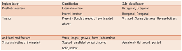
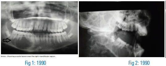
Endosseous Implants
Department of Prosthodontics A.B. Shetty Memorial Institute of Dental Sciences, Nitte University, Deralakatte, Mangalore, India
Abstract
Implants today are the most important and popular modality of replacing missing tooth /teeth. These osseointegrated supported prosthesis have overcome difficulties of retention which was previously experienced by a restorative prosthodontist in replacing missing teeth. This article reviews various guidelines as given in literature published by authors in English language from year 2000 to 2012. The key words used for the search were implant placement guidelines, implant angulations, single implant replacement, templates used to place implants, esthetics.
Key words: Endosseous Implants, Osseointegrated supported prosthesis, implant placement.
INTRODUCTION
Problems of complete denture retention and also problems of distal extension have been overcome by the use of implants. Esthetic regions with substantial bone loss was once considered to be replaced only by acrylic prosthesis to give the fullness for lip and check support caused due to loss of hard and soft tissues. In the recent years such assumptions are proved wrong by implant supported prostheses as they can overcome problems of retention and esthetics.
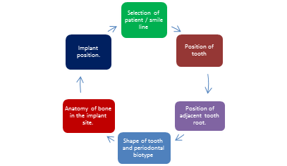
A successful implantologist is one who foresees the esthetic outcome even before placement of the implant by guiding the implants accurately into ideally in all the three dimensions namely the apicocoronal, buccolingual and mesiodistal dimensions for restoring the function, esthetics and comfort of the patient. Therefore implant placement should be prosthetically driven. A proper diagnosis and treatment planning along with establishing certain guidelines for implant placement are important requirements to achieve a favourable prosthetic outcome of the final prosthesis. This article reviews few articles published from 2000 to 2012 focusing on the guidelines for placement of oral implants to prevent crestal bone loss and provide a good emergence profile.
Classification of implant placement sites:

Factors to be considered in placement of implants in the anterior segment according to Jivraj and chee1
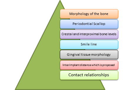
According to Becker2
Esthetics plays an important role in implants placed in the anterior maxilla 3. Also as the patient grows older the mandibular anterior teeth are more visible during speech and smile. Replacement of single-tooth with osseointegrated supported prosthesis maintains esthetics and helps to conserve dental tissues as well 4. Therefore placement and orientation of implant for ideal functioning and optimal esthetics is of prime importance in these regions 5.
Two important Guidelines for placing implants in the anterior region5
- Mesiodistal size of the natural crown to determine required space for the restoration
- Mesiodistal dimension of the root of the tooth which aids in determining the appropriate size of the implant.
Cemento Enamel Junction minus 2mm is the anatomic location which provides average size of tooth root and thus determines optimum size to be selected for implant to replace the missing tooth/teeth.
According to Babbush6, four parameters that help achievement of esthetic result and preservation of biologic health are:

Placement of implant in maxillary anterior region directly into the extraction socket should be avoided as this could lead to buccal plate perforation due to associated resorption that follows7.
Apicocoronal placement of implant:
Implant head should be placed a minimum of 3mm apical to a line that connects CEJ’s of adjacent teeth8. In case of existing recession, the implant head is to be placed 3mm from the gingival margins adjacent to the implant and 1.5-2mm from the CEJ of adjacent teeth in the absence of recession9. These apicocoronal placements will aid in masking the metal of implant and abutment which is also dependent on the design of the implant head. Superficial placement of implant results in visibility of metal margin and optical reflection due to bone loss around it. Therefore more apically the implant is placed better is the esthetics but poorer is the tissue health. Hence implant shoulder for a single stage implant should sink into a depth of 1-2mm and upto a depth of 2-3mm for a two stage implant apical to the imaginary line that joins the CEJ of the adjacent tooth8. This will help in developing an optimal gingival health as well as a proper emergence profile and for placement of porcelain of restoration subgingivally 6.
Mesiodistal implant positioning:
Mesiodistally the implant body should not be as wide as the natural tooth or the clinical crown it is intending to replace as this poses difficulty in establishing a proper emergence profile and maintaining the interdental papilla9. Ideally, the mesiodistal width of the implant should be equal to the contralateral tooth so that an optimal esthetic outcome can be achieved1. The distance from the adjacent tooth root or an implant also is a determinant of the implant diameter9.
Implant should be 1.5mm from the adjacent root ( Fig 1) to prevent wedge shaped vertical defect progressing to a horizontal defect resulting in bone loss of the adjacent tooth. Implants placed adjacent to each other should be spaced 3mm apart ( Fig 2) especially when one expects crestal bone loss . According to Carl Misch9 , the diameters of implants for replacing central incisors, lateral incisors and canines should be 5.2mm, 3.5mm and 4.2mm respectively1,6,7,8,9,10.
Buccolingual / Faciolingual position1, 7, 10, 11:
Single implant placed in maxillary anterior region should be placed slightly palatal to an imaginary line outlines which is formed by facial surfaces of the teeth adjacent to the implant. Too much of a palatal placement of implant causes development of unhygienic contours compromising biomechanics at the same time. Too much of buccal placement may end in dehiscence of buccal cortical plate and gingival recession in the area. 1-2 mm minimum thickness of buccal bone ( Fig 1) should thus be maintained to prevent recession and for improved esthetics.
Implant number and position:
Bone quantity and quality, force anticipated to be placed on restoration, relation of the arch form and shape of residual ridge determines the number of implants to be placed. In the less dense maxillary posterior region a total of three implants, one for each tooth being replaced is recommended. For fixed implant prosthesis with cantilever pontics, placement of six to eight implants is recommended in the maxilla. Additional implant placement is recommended with poor quality ridge and in case of bruxers where more force is anticipated on the implants12, 13. Dental arches with ovoid shape demand for three implants in the premaxilla in comparison to square dental arches which ask for only two implants since the central and lateral incisors are not cantilevered facially 13.
Four to six implants are the required to be placed between the mental foramina for hybrid prosthesis. Short implants can be placed distal to the mental foramina for long cantilever to reduce mechanical failure where the overdenture is not connected but merely rests on the implant to accommodate the flexion of mandible during the action of opening and closing of mandible 3.
Posterior placement of implant is sometimes contraindicated due to the presence of mental nerve loop situated 2 to 9 mm anterior to the mental foramina, in such cases posterior tilting of implants by 25-35°can be advantageous by reduction of cantilever length and better distribution of load, moves the implant support posterior, increases implant length, implant can follow dense bony structure and improves bony anchorage capacities in the edentulous mandible. In the edentulous maxilla, implants can be tilted 30-35° to avoid anterior wall of the sinus which has advantages similar to the implants placed in mandible and does not have any biological disadvantages 14.
Distribution of implant in the arch:
Implants should be spaced in the arch such that they share and distribute the occlusal loads equally, minimise cantilever distance and improve biomechanics. This distribution is of importance mainly in implant supported overdentures. Implant placement should be such that the bar constructed for overdenture is connecting implants in a straight line and prevents impingement of the denture bearing area and allow for adequate room for the clip 8, 13. Implant placement should be avoided in interproximal areas as they hinder with hygiene maintenance and compromise esthetics13.
Importance of Surgical guides:
High precision is required in placement of implants to achieve optimal esthetics, phonetics and function. A profile surgical template ( Fig 3) can be used successfully in completely edentulous maxilla with microimplants and transitional implants for stabilisation of template 5, 15. Templates were used with radiopaque markers, such as guttapercha, metal bearings, lead foil, metal rods or tubes resin teeth made with barium sulphate for proper dimension, location, and angulation of implant based on bone available, vital structures, and prosthesis predesigned16. Surgical templates can be tooth, bone and/ mucosa supported,Can be stereolithographic templates as well ( Fig 4 ). Implant Position Orthogonal Projection / IPOP validation method is supposed to be one of the accurate methods to evaluate the position of implant and detect inaccuracies of virtual implant planning systems. It evaluates the position of implant in the true buccolingual and mesiodistal positions in relation to the surrounding anatomical positions. It is also used to evaluate implant position after bone augmentation procedures and also for evaluation of extra oral implants17.
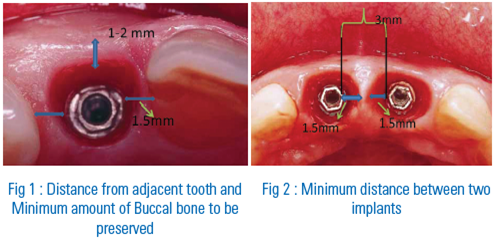
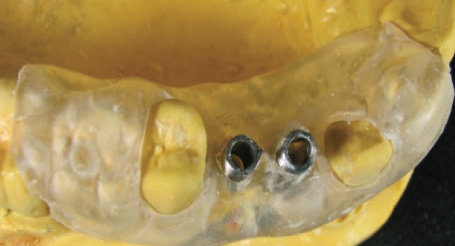
Fig 3: Surgical template

Fig 4: Stereolithographic template
DISCUSSION
Implants in the near future will be the most demanded treatment modality. Extreme high success rates, improved quality and ease of use and availability have thus attracted both practitioners and patients towards implant supported prosthesis as a treatment of choice. Peri-implant architecture thus takes a major share in the aesthetic setup of any implant-supported restoration.
A direct influence of esthetic implant positioning is seen on the soft tissue profile and appearance after prosthetic replacement. The more precisely the implant is positioned; the easier it will be to obtain a natural-looking, osseointegrated supported restoration in its soft tissue and hard tissue housing. Any gingival mismatch can be avoided with optimal implant positioning, which minimizes the need for further corrective surgeries and soft tissue reconstructions in the future.
Surgical template is also another key determining factor to the optimal implant position in three dimension, since the precise implant placement complements esthetics, function, clear patient phonetics, and easier maintenance of oral hygiene Thus, transfer of the information regarding the pre-determined position and angulation for the implant fixture from the study cast onto the surgical site becomes mandatory to allow a prosthetic driven implant placement protocol. A precisely fabricated surgical template or guide has an active role in executing the treatment plan at the first stage of surgery; it assists in the maintenance of a healthy natural biological space between the implant and the neighbouring roots. A three dimensional positioning surgical template could also be constructed to indicate the distance required to countersink the implant, which makes the third dimension achievable, and adds to the buccolingual and the mesiodistal dimensions.
The computer-milled surgical templates are also available, which provides a connection between the CT scan and the surgical template. These use reformatted CT data that combines with a three dimensional simulation of the implant position and produces a computer-milled surgical template. The simulated implant position will be created by softwares like SIM/Plant .The coordinates of these positions are transferred to a five-axis computer-controlled milling machine to create the appliance. Drill guide components are installed into the milled surgical template which aids in the direction of drilling procedure8, 15, 18.
CONCLUSION
The aim of dental surgeon is to reduce the amount of bending moments that can generate around supporting implant. Achievement of these criteria is by a careful treatment plan that selects the ideal location and number of implants to be placed based on the availability of hard tissue in the edentulous site and the possible resorption that could take place has to be anticipated in advance. The patient and the dental surgeon both enjoy the advantages of esthetic treatment with implants due to its increased retentive factors and advanced designs available. The ease of placement and availability has now proved osseointegrated supported prosthesis to be the treatment of choice. The knowledge of placement guidelines thus is a basic requirement to achieve this goal and makes the implantologist a successful practitioner to set a beautiful smile on the patients face enhancing confidence.
REFERENCES
- Jivraj S, Chee W. Treatment planning of implants in the aesthetic zone. Br Dent J.2006; 201: 77-89.
- Becker W. Immediate implant placement: treatment planning and surgical steps for successful outcomes. Br Dent J.2006; 201(4): 199-205
- Chee W, Jivraj S. Treatment planning of the edentulous mandible. Br Dent J.2006; 201(6): 337-347
- Prasad DA, Prasad DK. Effect of implant placement in growing adults on craniofacial development: A literature review. J Dent Implant 2012; 2:97-102.
- Aalam AA, Reshad M, Chee WW, Nowzari H. Surgical template stabilization with transitional implants in the treatment of edentulous mandible: A technical note. Int J Oral Maxillofac Implants 2005; 20(3):462—65.
- Babbush CA. Dental Implants: The Art and Science.Philadelphia, W.B.Saunders Co.2001;85-103.
- Al-Sabbagh M. Implants in the Esthetic Zone. Dent Clin North Am 2006;50: 391–407.
- Misch CE. Dental implant prosthetics. Mosby, 2005; 303-8.
- Misch CE. Contemporary Implant Dentistry. 2nd ed. St Louis: Mosby Inc 401-8.
- Barcelos MJ, Novaes Júnior AB, Conz MB, Harari ND, Vidigal Júnior GM. Diagnosis and Treatment of Extraction Sockets in Preparation for Implant Placement: Report of Three Cases. Braz Dent J. 2008; 19(2): 159-64.
- Handelsman M. Surgical guidelines for dental implant placement. Br Dent J.2006; 201: 139-52.
- Jivraj S,.Chee W. Treatment planning of implants in posterior quadrants. Br Dent J.2006; 201: 13–23.
- Jivraj S, Chee W, Corrado P. Treatment planning of the edentulous maxilla. Br Dent J.2006; 201: 261-79.
- Krekmanov L , Kahn M , Rangert B, Lindström H .Tilting of Posterior Mandibular and Maxillary Implants for Improved Prosthesis Support. Int J Oral Maxillofac Implants 2000;15:405–14.
- Sicilia A, Enrile FJ, Buitrago P, Zubizarreta J, Evaluation of the Precision Obtained with a Fixed Surgical Template in the Placement of Implants for Rehabilitation of the Completely Edentulous Maxilla: A Clinical Report. Int J Oral Maxillofac Implants 2000; 15:272–7.
- Cehreli MC, Sahin S, Fabrication of a Dual-Purpose Surgical Template for Correct Labiopalatal Positioning of Dental Implants. Int J Oral Maxillofac Implants 2000; 15:278–282.
- Verhamme LM, Meijer GJ, Boumans T, Schutyser F, Berge SJ , Maal TJJ. A clinically relevant validation method for implant placement after virtual planning. Clin. Oral. Implants. Res. 0, 2012 / 1–8.
- Becker CM, Kaiser DA. Surgical guide for dental implant placement. J Prosthet Dent 2000; 83(2): 248-251.
Disclaimer from Guident
The views and opinions expressed in articles published in Guident are solely those of the authors and do not necessarily reflect the views of the editors, the editorial board or the publisher, Ivory India. Guident does not endorse any product, technique or treatment described. Clinical content is intended for qualified dental and medical professionals and is not a substitute for individual clinical judgement. Guident, its editors and the publisher accept no liability for any consequence arising from the use of information published in the journal.
How to cite this article
Dr. Anupama Prasad, Dr. Krishna Prasad, Dr. Harshitha Alva. Endosseous Implants. Guident. 2013 Jun;6(8). Available from: https://guident.in/articles/implantology/endosseous-implants.html