A Review on Bisphosphonate Use and Implant Therapy : A Conundrum
Department of Prosthodontics, Royal Dental College, Kerala, India
ABSTRACT
Osteonecrosis of the jaw has been described in patients taking bisphosphonates after oral surgery procedures, including the placement of dental implants. This review is an update of the relationship between bisphosphonates and dental implants. Results obtained by different authors are compared, contrasting earlier studies where an improvement in implant osseointegration using bisphosphonates was observed, with ones where statistically significant differences were found. Currently, dental implants are contraindicated in patients being treated with intravenous bisphosphonates. All patients treated with bisphosphonates must have the risk of possible loss of implants and the risk of suffering a bony necrosis of the operated jaw explained to them, and give their informed consent prior to dental implant surgery.
INTRODUCTION
Bisphosphonates are a group of medications that have become increasingly and more widely used in the management of certain bone diseases . They have been approved for use by the U.S Food and Drug administration for the treatment of osteoporosis, which is very common, particularly in our ageing population. Metastatic bone disease , end stages of breast or prostate cancer , Paget’s disease , fibrous dysplasia , osteoporosis, cancers of the liver, lungs or kidney, multiple myeloma, hypercalcemia secondary to bony metastatic malignancy or hyperparathyroidism and certain other bone conditions warrant the use of bisphosphonates.1.
Surveys show that upto 190 million prescriptions of bisphoshonates are being written worldwide.2 According to Gutta and Louis3, the iv-bisphosphonates, zoledronate and pamidronate are being used in over 2.5 million patients worldwide.
It is generally accepted that bisphosphonates reduce osteoclastic activity in bone.The discovery of the possible association between the bisphosphonates and osteonecrosis of the jaws prompted both the United States Food and Drug Administration and Novartis, the manufacturer of bisphosphonates used in cancer chemotherapy, to issue a warning to health care professionals in September 2004.4,5 As an increasing proportion of the population is being prescribed oral bisphosphonates. It is inevitable that some of these patients may require implant therapy. Therefore the effects that these drugs may have on implant survival and success is very important.
HISTORY
Bisphosphonates have been known to chemists since the mid-19th century, with their first synthesis in Germany in 1865 They were first used in the textile, fertilizer and oil industries for the prevention of scaling in pipes because of their ability to inhibit calcium carbonate precipitation. The biological importance of bisphosphonates did not become apparent until the1960s, when Herbert Fleisch and colleagues discovered that inorganic pyrophosphates (PPi) in plasma and urine inhibited calcium phosphate dissolution and precipitation in vitro. Bisphosphonates were introduced in the mid-1990s as an alternative to hormone replacement therapies for treatment of osteoporosis. In 1996 Fosamax (alendronate, marketed by Merck) was the first bisphosphonate drug approved in the U.S. for the treatment of osteoporosis.6,7
In 2002 , serious side effects after surgical and dental procedures , in patients on bisphosphonate therapy, were documented. Multiple cases of bisphosphonate osteonecrosis, bisphosphonate avascular necrosis, bisphonateosteomyelitis and bis – phossy jaw were reported. The first case of Bisphosphonate-related Osteonecrosis of the Jaw (BRONJ) in oral and maxillofacial surgery was reported in 2003 by Marx and Ruggiero and involved the failure of osseointegrated dental implants .
CHEMISTRY AND MODE OF ACTION .
Bisphosphonates are synthetic compounds with a chemical structure similar to that of inorganic pyrophosphate, which is an endogenous regulator of bone mineralization.
Both oral BP and intravenous BP bond with bone and inhibit osteoclastic bone resorption, osteoclast activity,and induce their apoptosis .This is achieved by preventing the recruitment and function of osteoclasts. They also function indirectly by stimulating the osteoblasts to produce inhibitors of osteoclast formation. They can both decrease osteoclast activity and decrease osteoclast numbers. In high doses inhibition of ectopic and normal calcification can occur due to the physico-chemical inhibition of calcium phosphate crystal formation8. The most recent and powerful BP which include nitrogen in their molecule, inhibit tumor proliferation and angiogenesis
The half-life of bisphosphonates in the circulation is quite short, ranging from thirty minutes to two hours. However, once incorporated into bone tissue, they can persist for up to 10 years, depending on the skeletal turnover time.8,9. Of relevance to bone healing, Kapitola et al. reported bisphosphonates depress bone blood flow in rats and significantly decrease circulating levels of vascular endothelial growth factor which is important in the angiogenesis process.10
CLINICAL USES
One of the early clinical uses of bisphosphonates was as bone scan imaging agents. They remain very useful for detecting bone metastases and other bone lesions. Bisphosphonates are now used as the treatment of choice for inhibition of bone resorption in diseases where no effective treatment previously existed.Bisphosphonates effectively reduce the complications of the malignant osteolytic process. They reduce bone pain, the incidence of hypercalcemia and pathologic fracture, and the need for palliative radiotherapy. Overall, bisphosphonates improve the quality of life of patients suffering from abnormal bone resorption by reducing the number of pathological fractures and bone pain.
TYPES OF BISPHOSPHONATES
(11).
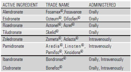
Currently, there are two main types of bisphosphonates: nitrogen containing and non-nitrogen containing.
A.Nitrogen containing bisphosphonates-
a. Oral- Alendronate (Fosamax) , Risedronate (Actonel) .
b. Intravenous - Pamidronate (Aredia, Pamisol) , Zoledronate (Zometa) .
B.Non- nitrogen containing bisphosphonates – Etidronate, Clodronate , Tiludronate .
Different Types Of Bisphosphonate In Current Usage.
-
BISPHOSPHONATE-RELATED OSTEONECROSIS OF THE JAW (BRONJ)
The Advisory Task Forces of the American Association of Oral and Maxillofacial Surgeons (AAOMS) and the American Society for Bone and Mineral Research (ASBMR), have defined BRONJ as :12
“Exposed, necrotic bone in the maxilla or mandible that has persisted for more than eight weeks, despite adequate treatment, in a patient with current or previous history of bisphosphonate use, without local evidence of malignancy and no prior radiotherapy to the affected region. Symptoms include delayed healing following a dental extraction or other oral surgery, pain, soft tissue infection and swelling, numbness, paraesthesia or exposed bone.
Symptoms include- pain, tooth mobility, mucosal swelling, and erythema. Common signs seen by a clinician may include parulis formation, sinus tract formation with or without active purulence, erythema, tooth mobility, or pain on palpation of affected tissue.Radiographs may appear normal or there may be widened lamina dura around teeth or ill-defined radiographic lytic lesions of bone with or without the evidence of sequestrum.These lesions can occur spontaneously or as a result of trauma, such as a surgical procedure.13,14
Wang et al.15 reported the first case of osteonecrosis of the jaw (ONJ) associated with bisphosphonate therapy in cancer patients. The majority of reported cases of bisphosphonates associated with ONJ have been diagnosed after invasive dental procedures such as tooth extraction16. Patients taking higher cumulative doses of bisphosphonates had greater risk of developing ONJ. ONJ is more prevalent in patients with malignancies than patients with benign bone disorders17.
Bisphosphonate-associated ONJ may range from a painless prolonged healing to a seriously painful and debilitating condition that lasts for many years without resolution. Treatment is difficult, and conventional surgical treatment or surgical curettage usually makes ONJ worse. Successful strategies include cessation of the bisphosphonate with symptomatic treatment with mouth rinses and antibiotics as indicated, and this may resolve the condition over many months or years.18
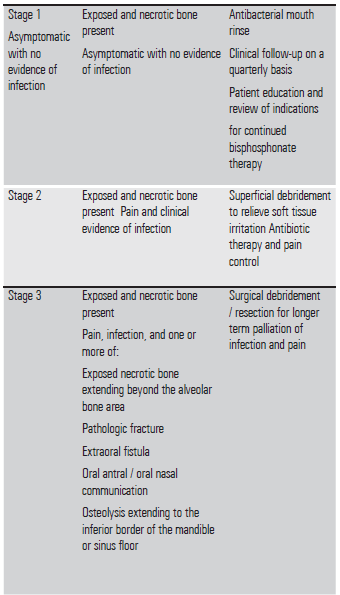
STAGING OF BRONJ
Current American Association of Oral and Maxillofacial Surgeons Guidelines on bisphosphonate-related osteonecrosis of the jaw Staging and Treatment (Ruggiero et al, 2009)

HISTOLOGY OF BRONJ
Favia et al. divided the BRONJ lesion into three major histological components19:
1.Areas of soft tissue composed of active inflammatory cells with acellular necrotic debris and bone resorption.
2. Areas with necrotic bony sequestra containing inflammatory cells.
3.Non-necrotic bone with increased trabecular thickness.
BISPHOSPHONATES AND DENTAL IMPLANTS
There have been several case reports and series that have evaluated implant failures in patients receiving intravenous bisphosphonate therapy (19).Other retrospective studies have also indicated that there may be risks of osteopathology developing in patients receiving oral bisphosphonates and dental implant treatment20-22.This is believed to be the result of reduced bone turnover due to diminished osteoclastic activity. There is mixed evidence regarding the association between oral bisphosphonate therapy and dental implant failure in the available literature related to this topic. Some studies found an association between the two, while other studies did not.
Grant et al.11 have described a series of 115 cases, of which eighty nine patients took bisphosphonates orally prior to implant surgery and thirty-three of them intravenously, for more than three years. No cases of osteonecrosis appeared and there were only two implants that failed to osseointegrate.Bell et al. obtained similar results published in an article that describes a series of 101 implants in forty-two patients; not a single case of osteonecrosis occurred. Of the sixty-one patients treated with 169 implants by Fugazzotto et al. with an average bisphosphonate medication period of 3.3 years, there was one case of bone exposure of the torus around a post-extraction implant, which closed spontaneously after four weeks.
BRONJ AND DENTAL IMPLANTS - HISTOLOGY
Kwon et al.19 conducted studies into the histological picture present in the BRONJ lesions that affected dental implant. The authors proposed three distinct patterns of BRONJ associated with dental implants:
- A “Frozen type”, in which there is abundant necrotic bone around the implant with little soft tissue.
- An “osteolytic type”, with extensive osteolysis around the implant with/without sequestrum formation. Typically, there is a combination of soft tissue that is clearly inflamed with residual necrotic and viable bone.
An “en block” type sequestration with the dental implant in the sequestrum. In this type of lesion there is still a considerable amount of bone-implant contact maintained. This “en block” sequestration has been seen in other studies involving the catastrophic failure of implants in bisphosphonate patients24-26.

FIGURE 2- BRONJ associated with dental implant. (A) Exposed necrotic bone area became to be infected and bone destruction continues outside of implant surrounding bone and maintaining the bone–implant contact. The lesion became to be sequestrated as a block including the implant, which could be termed “en block sequestration.” (B) When the alveolar bone around implant is totally necrotic and the inflammatory infiltration proceeds along with the implant surface, the implant might finally lose stability. The further osteolysis around previous implant site can result in the sequestra formation. These findings can co-exist according to the severity of the BRONJ.
LABORATORY TESTS
The CTx test ( C- terminal telopeptide and collagen type 1 C telopeptide ) is a serum blood test to monitor the markers of bone turnover and evaluate the risk of developing BRONJ. C-Telopeptides are fragments of collagen released during bone remodelling. Bisphosphonates reduce bone level thereby decreasing the serum CTx levels. Serum CTX can identify changes in bone remodelling within several days to 2 weeks.
Laboratory risk assessment –
cTx value (pg/ml) Risk for osteonecrosis
300- 600(normal) None.
150-299 None to minimal.
101- 149 Moderate .
< 100 High.
THE MARX PROTOCOL.
The protocol suggested by Marx (27) for dental implant candidates on bisphosphonates-
Oral bisphosphonate use > 3 years
- Physician’s approval to discontinue bisphosphonate 3 months before surgery and three months after surgery – Drug Holiday.
- CTx levels > 150pg/ml before planning implant surgery.
- Detailed informed consent.
Oral bisphosphonate use < 3 years without clinical or radiographic risk factors
- CTX levels must be > 150pg/ml.
- Proceed with surgery after informed consent for BRONJ.
- If CTx <150pg/ml- physician approved drug holiday and monitor every three months till CTx levels are >150pg/ml.
Oral bisphosphonate use < 3 years with clinical or radiographic risk factors
- Drug holiday for 3 months.
- If CTx <150pg/ml- physician approved drug holiday and monitor every three months till CTx levels are >150pg/ml.
- CTx levels must be > 150 pg/ml to proceed with informed consent for BRONJ.
DENTAL IMPLANT IMPLICATIONS.
In 2009, the American Association of Oral and Maxillofacial Surgeons (AAOMS ) formed a Task Force on Bisphosphonate- Related Osteonecrosis of the Jaws lead by Salvatore Ruggiero16.With respect to oral surgical treatment, this position paper clearly states that for patients who have been taking oral bisphosphonates for less than 3 years with no clinical risk factors, no alteration or delay in proposed surgery is required. Importantly, Ruggiero et al. recommends that if dental implants have been placed, then informed consent should be obtained from the patient outlining the risk of possible future implant failure and the risk of development of BRONJ.
The principles of oral surgery for the prevention of bisphosphonate-related osteonecrosis of the jaw outlined by Heufelder et al. and adopted by the department of Oral and Maxillofacial Surgery and Facial Plastic Surgery of the University of Leipzig can be modified to apply to implant patients on bisphosphonates.
- Perioperative oral antibiotics for 7 days, starting 48 hours before surgical intervention. All patients received the oral antibiotics amoxicillin and clavulanic acid (875 mg/125 mg) every 12 hours or, in cases of penicillin allergy, clindamycin 600 mg every 8 hours.
- Wound seal must be achieved using horizontal mattress sutures and single knots to adapt the edges of epithelium.
- Patients must be followed up 1, 2, 4, and 12 weeks and 6 and 12 months postsurgery.
- Antiseptic mouth rinsing, using 1% peroxide prescribed 3 times a day.
- Removal of the sutures must not performed until at least 14 days after surgery.
- Dental prostheses must be removed, strictly, for at least 3 weeks postsurgery
- Atraumatic approach - flapless guided implant placement in these patients.
- Prolonged healing times following surgery.
- Meticulous oral hygiene measures and maintenance schedules must be enforced.
- Recording of baseline levels of implant stability (ISQ) using RFA frequently to detect early loss of osseointegration in the absence of symptoms.
DISCUSSION
As with most laboratory tests, there are shortcomings associated with the CTX test. Serum CTX is known to be subject to circadian variation and can also be affected by fasting .CTX to determine treatment protocols for patients being treated with oral bisphosphonates has received much criticism and the reliability is questionable.Extensive research by Abtahi et al.32 showed that a bisphosphonate coating on a dental implant improved the fixation of dental implants in humans. They carried out a randomized double-blind study, in which fibrinogen-coated dental implants coated with pamidronate and ibandronate were inserted into the maxilla. The implant stability was determined by resonance frequency analysis (RFA) and implant stability quotient (ISQ) readings. Radiographic appearances were improved when compared to a control group. The theory behind this positive effect is that once bisphosphonates are released from the surface of the coated implant, there is a localized reduction in osteoclastic activity, altering the balance of bone remodelling in favour of increased osteoblastic activity, leading to increased bone formation which in turn increases the local bone density32,33. The authors hope that this improved early fixation may reduce the rate of late implant failures and could also allow earlier loading times.
CONCLUSION
Most studies show that there is a certain risk associated with the both implant placement and the maintanence of osseointegrated implants in patients receiving oral bisphosphonates. Bisphosphonate related implant failure appears to be more common in women than in men. This may be due to the fact that a greater proportion of the population receiving bisphosphonates tends to be post-menopausal osteoporotic women. The duration of bisphosphonate therapy is critically important with respect to the development of BRONJ. The posterior regions of both the maxilla and mandible appear to be sites most frequently affected with respect to implant failure. Those cases that affect the posterior maxilla are possibly due to an already existing poor quality of bone which is further diminishedby the reduction in bone turnover caused by bisphosphonate. Late implant failures appear to occur in patients treated with oral bisphosphonate exposure for a period >3 years especially in
patients who have existing integrated implants10, while early failures appear to occur in patients treated with bisphosphonates before or at the time of implant placement. The utility of bone turnover markers in guiding oral surgical treatment decisions is controversial
therapy. These conclusions demand further investigations with significantly larger populations over longer periods of bisphosphonate treatment exposure. Moreover, novel strategies for the prevention, risk reduction, and treatment of BRONJ need to be developed further so that more accurate judgments about risk, prognosis, treatment selection, and outcome can be established for patients with BRONJ.
REFERENCES
1. A Cheng, A Ma vrokokki, G Carter, B Stein, NL Fazzalari, DF Wilson, AN Goss.The dental implications of bisphosphonates and bone disease. Australian Dental Journal Medications Supplement 2005; 50:4.
2.Elliott Ballantyne .Bisphosphonates: Possible Modes of Action and Implications for Dental Implant Treatment. A Review of the Literature . Journal of General Practice. Volume 3 • Issue 1 .
3.Gutta R, Louis PJ . Bisphosphonates and osteonecrosis of the jaws:
science and rationale. Oral Surgery Oral Medicine Oral Pathology Oral Radiology Endodtics . 2007 104: 186-193.
4. Ravosa MJ, Ning J, Liu Y, Stack MS (2011) Bisphosphonate effects on the behaviour of oral epithelial cells and oral fibroblasts. Archives Of Oral Biology 56: 491-498.
5. Kharazmi M, Persson U, Warfvinge G (2012) Pharmacovigilance of oral
bisphosphonates: adverse effects manifesting in the soft tissue of the oral
cavity. Journal Of Oral And Maxillofacial Surgery. 70: 2793-2797.
6. Fleisch H. Development of bisphosphonates. Breast Cancer Research . 2002; 4:30-34.
7. Antonio-Juan Flichy-Fernández, José Balaguer-Martínez, Miguel Peñarrocha-Diago, José V. Bagán. Bisphosphonates and dental implants: Current problems .Medicina Oral Patologia Oral YCircugia Bucal. 2009 Jul 1;14 (7):E355-60.
8. Martin TJ, Grin V. Bisphosphonates – mechanisms of action. Australian Prescriber 2000;23:130-132.
9.Sparidans RW, Twiss IM, Talbot S. Bisphosphonates in bone diseases. Pharm World Sci 1998;20:206-213.
10. Wood J, Bonjean K, Ruetz S, et al. Novel antiangiogenic effects of the bisphosphonate compound zoledronic acid. J Pharmacol Exp Ther 2002;302:1055-1061.
11.José-M,Montoya-Carralero , Pablo Parra-Mino , Piedad Ramírez-Fernández , Isabel Mª Morata-Murcia , Mª del Carmen Mompeán-Gambín , José-Luis Calvo-Guirado.Dental implants in patients treated with oral bisphosphonates. A bibliographic review . Medicina Oral Patologia Oral YCirugia Bucal. 2010 Jan 1;15 (1):e65-9.
12. Alastair Goss, Mark Bartold, Paul Sambrook, and Peter Hawker .The Nature and Frequency of Bisphosphonate-Associated Osteonecrosis of the Jaws in Dental Implant Patients: A South Australian Case Series. Journal Of Oral Maxillofacial Surgery 68:337-343, 2010
13. S Patel, S Choyee, J Uyanne, AL Nguyen, P Lee, PP Sedghizadeh, SKS Kumar, J Lytle,S Shi, AD Le. Non-exposed bisphosphonate-related osteonecrosis of the jaw: a critical assessment of current definition, staging, and treatment guidelines . Oral Diseases (2012) 18, 625–632
14.Vudiniabola S, Pirone C, Williamson J, Goss AN. Hyperbaric oxygen in the therapeutic management of osteoradionecrosis of the facial bones. International Journal Of Oral And Maxillofacial Surgery. 2000;29:435-438.
15. Wang EP, Kaban LB, Streler GJ, Raje N, Troulis MJ .Incidence of osteonecrosis of the jaw in patients with multiple myeloma and breast ,prostate cancer on intravenous bisphosphonate therapy. Journal Of Oral And Maxillofacial Surgery. 2003. 65: 1328-1331.
16. Ruggiero SL, Mehrotra B, Rosenberg TJ, Engroff SL (2004) Osteonecrosis of the jaws associated with the use of bisphosphonates: a review of 63 cases. J Oral Maxillofac Surg 62: 527-534.
17. Hoff AO, Toth B, Hu M, Hortobagyi GN, Gagel RF .Epidemiology and risk factors for osteonecrosis of the jaw in cancer patients. Annals Of New York Academy Of Sciences . 2011.1218: 47-54.
18.Cheng A, Daly CG, Logan RL, et al: Alveolar bone and the bisphosphonates. Australian Dental Journal. 54:S51, 2009
19 .Kwon TG, Lee CO, Park JW, Choi SY, Rijal G, et al. Osteonecrosis associated with dental implants in patients undergoing bisphosphonate treatment.Clinical Oral Implants Research.2014. 25: 632-640.
20.Ayan M, Dolanmaz D, Mihmanlı A, Ayan A, Karkasam. (2012) The effect of systemically administrated zoledronic acid on the osseointegration of dental implants.Oral Dis 18: 802-808.
21.Martin DC, O'Ryan FS, Indresano AT, Bogdanos P, Wang B, et al. (2010) Characteristics of implant failures in patients with a history of oral bisphosphonate therapy.Journal Of Oral And Maxillofacial Surgery 68: 508-514.
22.Yip JK, Borrell LN, Cho SC, Francisco H, Tarnow DP (2012) Association between oral bisphosphonate use and dental implant failure among middle-aged women.Journal Of Clinical Periodontology 39: 408-414.
23. Favia G, Pilolli GPMaiorano E. Histologic and histomorphometric features of bisphosphonate-related osteonecrosis of the jaws: an analysis of 31 cases with confocal laser scanning microscopy. Bone , 2009.406-13.
24. Yuan K, Chen KC, Chan YJ, Tsai CC, Chen HH, et al. (2012) Dental implant failure associated with bacterial infection and long-term bisphosphonate usage: a case report.Implant Dent 21: 3-7.
25. Shirota T, Nakamura A, Matsui Y, Hatori M, Nakamura M, et al. (2009) Bisphosphonate-related osteonecrosis of the jaw around dental implants in the maxilla: report of a case.Clin Oral Implants Res 20: 1402-1408.
26. Lazarovici TS, Yahalom R, Taicher S, Schwartz-Arad D, Peleg O, et al. (2010) Bisphosphonate-related osteonecrosis of the jaw associated with dental implants.J Oral MaxillofacSurg 68: 790-796.
27. Marx RE . Pamidronate (Aredia) and zoledronate (Zometa) induced avascular necrosis of the jaws: a growing epidemic. Journal Oral And Maxillofacial Surgery 61: 2003,1115-1117
28. Marx R (2007) Oral and Intravenous Bisphosphonate-Induced Osteonecrosis of the Jaws: History, Eitiology, Prevention and Treatment. Quintessence Publishing Co, Inc
29.Woo SB, Kalmar JR (2007) Osteonecrosis of the jaws and bisphosphonates. Alpha Omegan 100: 194-202.
30. Ott SM (2011) What is the optimal duration of bisphosphonate therapy? Cleveland Clinical Journal Of Medicine 78: 619-630.
31. McLeod NM, Brennan PA, Ruggiero SL (2012) Bisphosphonate osteonecrosis of the jaw: a historical and contemporary review. Surgeon 10: 36-42.
32. Abtahi J, Tengvall P, Aspenberg P (2012) A bisphosphonate-coating improves the fixation of metal implants in human bone. A randomized trial of dental implants. Bone 50: 1148-1151.
33. Berardi D, Carlesi T, Rossi F, Calderini M, Volpi R, et al. (2007) Potential applications of biphosphonates in dental surgical implants. Int J Immunopathol Pharmacol 20: 455-465.
Disclaimer from Guident
The views and opinions expressed in articles published in Guident are solely those of the authors and do not necessarily reflect the views of the editors, the editorial board or the publisher, Ivory India. Guident does not endorse any product, technique or treatment described. Clinical content is intended for qualified dental and medical professionals and is not a substitute for individual clinical judgement. Guident, its editors and the publisher accept no liability for any consequence arising from the use of information published in the journal.
How to cite this article
Dr. Shajahan PA, Dr. Rohit Raghavan, Dr Sumitra S. A Review on Bisphosphonate Use and Implant Therapy : A Conundrum. Guident. 2016 May;9(6). Available from: https://guident.in/articles/implantology/a-review-on-bisphosphonate-use-and-implant-therapy-a-conundrum.html