A Combined Approach to Restore a Fractured Tooth: Reattachment and Composite Veneering
Department of Periodontist & Implantologist, Christian Dental College, CMC&H, Ludhiana, Punjab, India
ABSTRACT
Traumatic dental injuries are a common occurrence. Numerous treatment options are available for restoring such a tooth. Careful examination of tooth, tooth fragments and fracture line are important aspects before finalising treatment plan. Meticulous and scientific approach can sometimes guide you to an innovative treatment plan. This paper is an attempt to report one such case. An adult with trauma to his central incisor, reported with coronal fracture. Fracture lead to three fragments; two palatal and one facial. Patient had preserved only the palatal fracture fragments. A treatment option was selected which gave the patient natural palatal aspect and artificial facial aspect. The treatment selected was based on the fact that fracture line was favourable. Also, natural contour, texture and wear rate could be maintained for the palatal aspect. The facial side was cut like a veneer preparation and restored with composite. This gave an advantage of natural mesial, distal contour and perfect matching aesthetic composite on facial aspect. Patient was pleased with the aesthetic result and emotionally benefited from the fact that his natural tooth fragments were used.
KEY WORDS: Crown fracture, veneering, reattachment, composite.
INTRODUCTION
Dental trauma is a prevalent condition and coronal fracture of anterior teeth being the most common1. It is not only a cause of discomfort to patient but also effect patient’s confidence .Periodontal, endodontic, coronal and occlusal assessment of such a case is vital for treatment planning2. With no or minimal biological width violation and fractured fragment in good condition, fragment reattachment is a good treatment choice3. Especially with advancements in bonding agents, this simple and low cost technique is currently preferred. Fractured fragment reattachment with facial veneering is a better option to a complete composite restoration, providing advantages like natural wear, contour, texture and better masticatory function on occluding side.
CASE REPORT
A male patient aged 47 years reported to the Department of Conservative Dentistry and Endodontic, Christian Dental College, CMC&H, Ludhiana, India with the chief complaint of broken upper front tooth. History revealed that the tooth fractured 5 days back when the patient, who is a factory worker, was trying to open some tool with his teeth. He did not avail any dental treatment after the episode.
Clinical examination revealed fractured upper left central incisor (Figure 1, 2). There was no clinical exposure of pulp (Figure 3). Fracture line extended from incisofacial to gingivopalatal direction. Patient had two palatal fragments of teeth with him, wrapped in a piece of paper (Figure 4). On examination of fractured fragments three observations were made. The first observation was that the fragments adapted well with each other and to the remaining tooth structure. Secondly, that the colour of the fragments did not match with the remaining crown structure. And thirdly, a small fractured fragment was missing from the facial aspect.
Patient complained of sensitivity to cold since the day of trauma. Pain was transient and relieved immediately after removal of stimulus. Thermal test was conducted on fractured and contra lateral tooth. It revealed early response in 21 as compared to 11. Electric pulp test showed similar results. Patient was explained all treatment options. Considering patients inclination for reattachment of fractured fragment, informed consent for the treatment was taken.
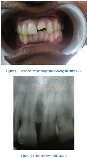

Figure (3): Palatal view of 21.
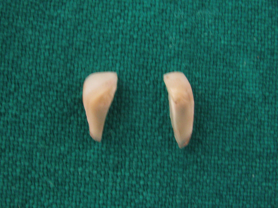
Figure (4): Palatal fragments.
The fractured fragments were rehydrated by immersing in normal physiological saline solution. The two palatal fragments were then etched using 37% phosphoric acid for 15 seconds and washed for 30 seconds (Figure 5). Bonding agent Tetric N Bond (Ivoclar Vivadent) was applied on both the fragments, rubbed for 10 seconds, gently air thinned and light cured for 10 seconds. The two fractured fragments were then united using flowable composite Tetric N Flow (Ivoclar Vivadent), which was light cured for 20 seconds. Excess material which could hinder adaptation of the united fragment to remaining tooth structure was meticulously removed using diamond bur.
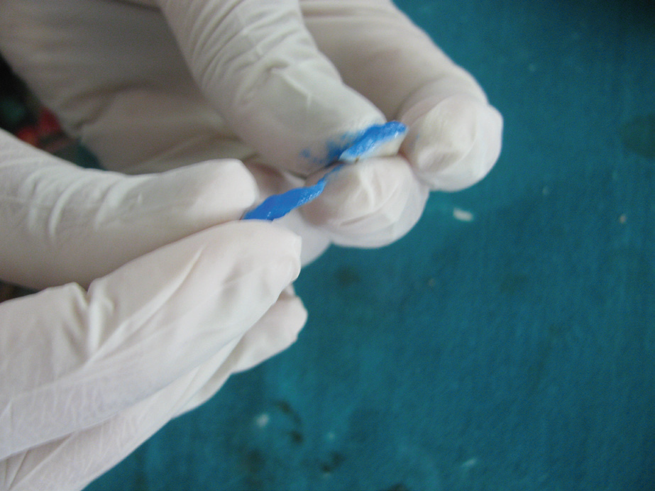
Figure (5): Acid etching of palatal fragments.
The fused palatal fragments were then checked for adaptation to remaining tooth (Figure 6). Thereafter, the united palatal fragments and the remaining tooth structure were bonded using the same method as mentioned above. After reattachment, facial aspect was prepared like a veneer preparation (Figure 7). A Light cured composite shade A2 (Z-100,3M ESPE) was used to restore the facial aspect (Figure 8). Composite veneer was finished using Sof-lex discs (3M ESPE). After restoration, occlusion was checked to avoid any premature contact. Patient was recalled after 4weeks. Tooth was functional and asymptomatic (Figure 9). Electric pulp testing and thermal test gave normal results.
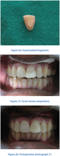

Figure (9): Palatal View of 21.
DISCUSSION
Various treatment options are available with the dentist for treating a case of coronal fracture. These include; fracture reattachment, composite buildup or a full coverage crown. The treatment approach adopted depends upon location of fracture, extent of fracture and adaptability of fractured fragments. Finally, it is also the choice made by the patient after considering all pros and cons.
In this case, patient opted for fractured fragment reattachment with facial veneering. Patient had two palatal fractured fragments which adapted well with each other and to the remaining tooth structure. The fragments revealed shallow knife edged subgingival fracture margin which minimally invaded biological width and did not require any special intervention4. When adapted to the tooth, these fragments reproduced palatal contour of the tooth beautifully.
From the facial aspect, a small fracture fragment was missing. Also, there was lack of colour match between remaining tooth and fractured fragments. Composite veneering was planned for the facial aspect. Facial veneering not only served the purpose of aesthetics, but it also increased the resistance to shear stresses at the fracture line1 (Figure 10). Other features like bevelling and chamfer preparation have also been proposed for enhancing resistance to fracture reattachment5.
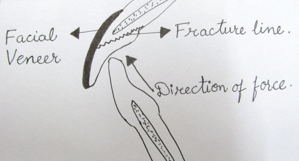
Figure (10): Diagrammatic representation of treatment.
The palatal side had reattached palatal fragments. This provided natural wear as compared to composite restoration(6) and maintained incisal guide in tooth structure. Reattachment also established natural contour, texture and positive psychological response(7). It is a simple and conservative approach which is especially recommended for young patients.
Reports and clinical experience indicate that the reattachment has a successful medium term outcome8, 9. Patients should be informed about the interim nature of the treatment and that it is an alternative to a more invasive prosthetic treatment10.
REFERENCES
- Hamilton FA, Hill FJ, Holloway PJ. An investigation of dento-alveolar trauma and its treatment in an adolescent population. Part 1: The prevalence and incidence of injuries and the extent and adequacy of treatment received. Br Dent J 1997; 182:91-95.
- Chu CJF and Wei SHY. Clinical consideration for fracture reattachment. Quintessence Int 2000; 31:385-391.
- Baratieri LN, Ritter AV, Monteiro S Jr, de Mello Filho JC. Tooth fracture reattachment: An alternative for restoration of fractured anterior teeth. Pract periodontics Aesthet Dent 1998;10:115-125.
- Macedo GV, Diaz PI, de O Fernandes CA, Ritter AV. Reattachment of anterior teeth fragments: A conservative approach. J Esthet Restor Dent 2008; 20:5-20.
- Reis A, Loguercio AD, Kraul A, Matson E. Reattachment of fractured teeth: A review of literature regarding techniques and materials. Oper Dent 2004; 29:226-233.
- Baratieri LN, Monteiro S Jr, Andrada MA. Tooth fracture reattachment a case report. Quintessence Int1990; 21:261-270.
- Maia EA, Baratieri LN, de Andrada MA. Tooth fracture reattachment: Fundamentals of techniques and two case reports. Quintessence Int 2003; 34:99-107.
- Andreasen FM, Noren JG, Andreasen JO. Long term survival of fragment bonding in the treatment of fractured crowns. Quintessence Int 1995; 26:669-681.
- Oz IA, Haytac MC, Toroglu MS. Multidisciplinary approach to the rehabilitation of a crown root fracture with original segment for immediate aesthetics: case report with 4 –year follow up. Dent Traumatol 2006; 22:48-52.
- Sharma D, Garg S, Sheoran N, Swami S, Singh G. Multidisciplinary approach to the rehabilitation of a tooth with two trauma episodes: Systemic review and a case report. Dent Traumatol 2011; 27:321-326.
Disclaimer from Guident
The views and opinions expressed in articles published in Guident are solely those of the authors and do not necessarily reflect the views of the editors, the editorial board or the publisher, Ivory India. Guident does not endorse any product, technique or treatment described. Clinical content is intended for qualified dental and medical professionals and is not a substitute for individual clinical judgement. Guident, its editors and the publisher accept no liability for any consequence arising from the use of information published in the journal.
How to cite this article
Dr.Preet Kanwal Kaur Atwal Department of Conservative Dentistry and Endodontics, Dr. Rajbir Singh. A Combined Approach to Restore a Fractured Tooth: Reattachment and Composite Veneering. Guident. 2016 May;9(6). Available from: https://guident.in/articles/endodontics/a-combined-approach-to-restore-a-fractured-tooth-reattachment-and-composite-veneering.html