Management of Large Radicular Cyst
Department of Conservative Dentistry & Endodontics, Department of Oral & Maxillofcial Surgery, Seema Dental College & Hospital, Rishikesh, Uttrakhand, India
Abstract
Radicular cysts are the most common odontogenic cystic lesions of inflammatory origin and are managed either by surgical enucleation or by marsupialization. This article aims to report a clinical case of radicular cyst treated with surgical enucleation.
INTRODUCTION
The most frequent odontogenic cyst of teeth bearing areas is the radicular cyst, also called periapical cyst. Two techniques can be used in clinical practice for surgical removal of cysts :enucleation and marsupialization.1
Enucleation is defined as a complete removal of the cystic lining with healing by primary intention while marsupialization is synonymous with Partsch’s operation, and is the conversion of a cyst into a pouch.2
Although small cystic lesions frequently heal simply with endodontic therapy, larger lesions may need additional treatment. Untreated cysts may expand causing local tissue destruction and deformities.1
This article aims to report a clinical case of radicular cyst treated with surgical enucleation.
CASE REPORT
A 50 year old female patient reported to the Department of Conservative Dentistry and Endodontics, Seema Dental College and Hospital, Rishikesh with a painless swelling in the upper front tooth region (Fig 1).

Figure 1
The patient was unable to describe precisely how long the lesion had been present. The lesion was painless and there was no history of epistaxis or nasal discharge, although the patient reported partial obstruction of the nasal cavity. Extraoral examination revealed facial asymmetry. On palpation, the lesion was nodular, soft, fluctuant and circumscribed.
Intraoral examination revealed presence of a nodular lesion consistent with the extraoral findings, extending anteriorly toward the alveololabial groove. The mass was rounded, swollen, and clearly circumscribed(Fig 2).
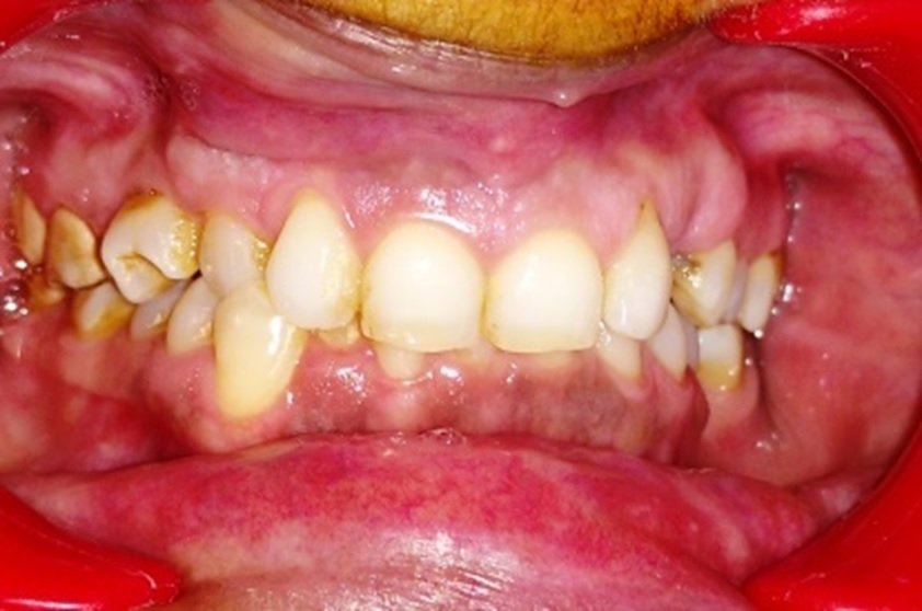
Figure 2
The patient had no systemic comorbidities.
On radiographic examination it was seen that the cyst was bilateral, involving teeth from the upper right canine to the upper left lateral incisor(Fig 3).

Figure 3
On vitality testing, the teeth were non vital. The teeth were then treated endodontically, in which access preparation and cleaning and shaping upto Protaper F2 were carried pre-operatively(Fig 4, 5).
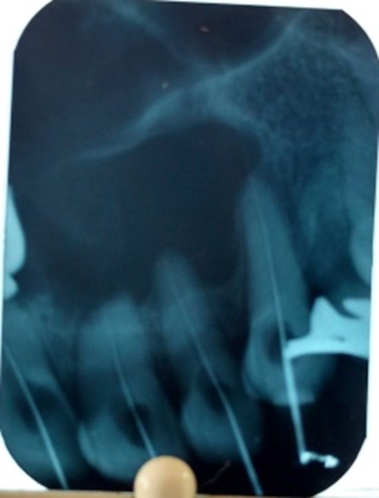
Figure 4

Figure 5
For the surgical approach, left and right sided infraorbital and nasopalatine nerve blocks were administered, a trapezoidal flap was raised in which the vertical releasing incisions were given corresponding to the first premolar region of either side(Fig 6).

Figure 6
On raising the flap it was seen that the cyst was secondarily infected(Fig 7),

Figure 7
as there was presence of exudate, which was aspirated(Fig 8).

Figure 8
The cyst was enucleated using curettes and periosteal elevators(Fig 9).

Figure 9
After thorough cleaning and debridement of the cavity, the teeth were obturated using F2 GP and root canal sealer(Fig 10).
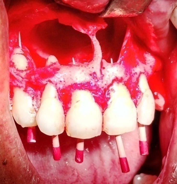
Figure 10
During the enucleation, it was observed that the nasal floor was perforated by the cyst. To help in post operative healing, the cavity was packed with gel foam(Fig 11).
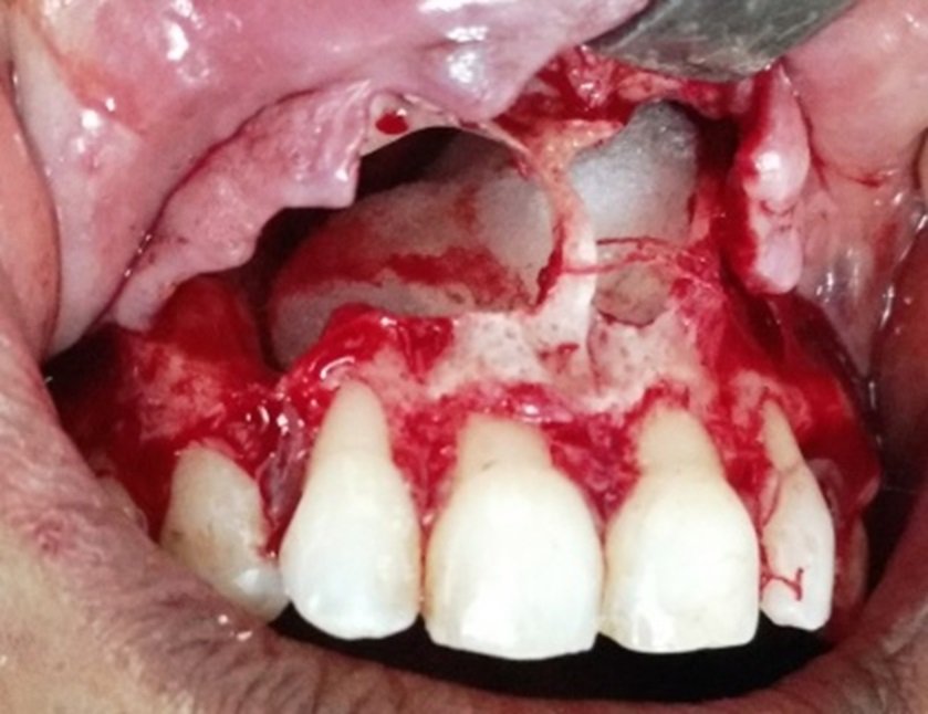
Figure 11
The soft tissues were then pulled back and sutured using sling sutures which were removed on the 7th postoperative day(Fig 12).
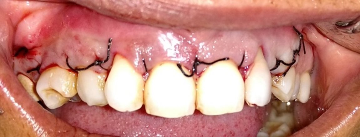
Figure 12
DISCUSSION
The surgical approach to cystic lesions of the jaws is either marsupialization or enucleation.3The treatment of choice is dependent on the size and localization of the lesion, the bone integrity of the cystic wall and its proximity to vital structures. Cysts are usually enucleated, where the cystic lining is separated from its inner bony surface and removed and the cavity allowed filling with blood clot.
The treatments of cysts are still under discussion and many professionals option for a conservative treatment by means of endodontic technique. However in large lesions the endodontic treatment alone is not efficient and it should be associated to a decompression or a marsupialization or even to enucleation.1
Post-surgical period was uneventful. The cystic lesion was submitted for histopathological examination. Histopathological features were consistent with the clinical diagnosis of infected radicular cyst. The choice of treatment may be determined by somefactors such as the extension of the lesion, relation with noble structures, evolution, origin, clinical characteristic of the lesion, cooperation and systemic condition of the patient.4
CONCLUSION
The clinical case reported in this article was managed successfully by endodontic therapy followed by surgery. It is suggested that the treatment of the radicular cysts should be defined according to the clinical and radiographic evaluations according to each case.
REFERENCES
- FawziRiachi, CarineTabarani. Effective Management of Large Radicular Cysts Using Surgical Enucleationvs Marsupialization Two Case Reports. IAJD; 1(1):44-51
- Neaverth EJ, Burg HA.Decompression of large periapical cystic lesions. J Endodont1982 ; 8 : 175-182
- Bodner L. Cystic lesions of the jaws in children.Int J PediatOtorhinolaryngol 2002; 62: 25-29.
- Narendra.U. Manwar, Abhishek Agrawal, Manoj.G. Chandak. Management of infected radicular cyst by surgical approach. International journal of dental clinics 2011:3(4):75-76.
Disclaimer from Guident
The views and opinions expressed in articles published in Guident are solely those of the authors and do not necessarily reflect the views of the editors, the editorial board or the publisher, Ivory India. Guident does not endorse any product, technique or treatment described. Clinical content is intended for qualified dental and medical professionals and is not a substitute for individual clinical judgement. Guident, its editors and the publisher accept no liability for any consequence arising from the use of information published in the journal.
How to cite this article
Dr. Bharatendu Kawatra, Dr. Philip Pradeep, Dr. R. Muthunagai. Management of Large Radicular Cyst. Guident. 2016 Jun;9(7). Available from: https://guident.in/articles/endodontics/management-of-large-radicular-cyst.html