Oral Mucocele: LASER Excision Using Diode LASER-940NM
Urbana Dental Clinic & LASER Centre, Faridabad, Haryana
ABSTRACT
The “mucus filled cavities” that occur in the oral cavity mainly as a result of mechanical trauma that leads to rupture of salivary gland ductal system and causes mucin spillage into the adjacent oral soft tissues. These may interfere with mastication, speech and esthetics. The present case report illustrates excision of mucocele using 940 nm Diode Laser.
INTRODUCTION
Mucoceles are the “mucus filled cavities” present in various cavities such as oral cavity, paranasal sinuses and lacrimal sac.1 Mechanical obstruction of salivary glands results in salivary obstructive diseases; mucus extravasation and mucus retention being the most common types of diseases of which mechanical obstruction is the main cause.2 Extravasation cyst occurs due to rupture of the salivary gland ductal system as a result of mechanical trauma causing spillage of mucin into the adjacent soft tissues.3,4 Mucus retention cysts occur markedly by the obstruction of salivary ductal walls that results in dilatation of the salivary ducts without spillage of any mucin.5,6
CASE REPORT
A 6 years old female patient presented a swelling in the labial mucosa of left lower region. Swelling had been persisting for few months and also interfered in mastication (Figure 1).
Clinical examination revealed a soft, painless swelling which was filled with fluid. The lesion measured approximately 1 × 1 cm in size. Clinical examination and history of the lesion presented by the patient concluded the lesion to be Mucocele.
There are many approaches for treatment of mucoceles which were explained to the patient’s guardian and an informed written consent form was acquired from her father for the excision of the lesion with the most recent treatment modality available, LASER.
Marginal infiltration with 1:80,000 Xylocaine was done and removal was carried out using soft tissue 940 nm Diode LASER, diameter of the tip was 400µm with power setting of 1.5W under continuous mode (Figure 2). Comprehensive excision of the mucocele was done using undermining strokes (Figure 3,4). The specimen was exposed to histopathological analysis (Figure 6,7). Patient was prescribed analgesics.

Figure 1. Pre-operative
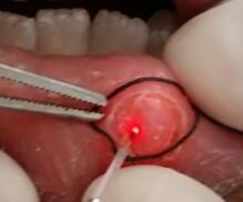
Figure 2. Intra-operative: Mucocele circumscribed with suture
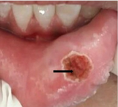
Figure 3. Immediately after excision of Mucocele
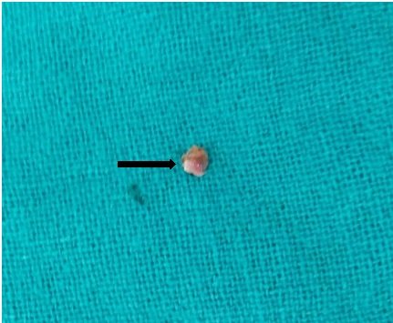
Figure 4. Tissue excised for histopathological examination

Figure 5. Post-operative after 15 days

Figure 6. Histopathology-Mucin filled space & Figure 7. Mucinophages
DISCUSSION
Mucoceles are among the most common oral lesions. Mucoceles are more prevalent in children and young adults with an equal or no predilection for any gender.7 Mucoceles are dome-shaped elevated swellings that accumulate mucin and is the characteristic feature of mucocele. These lesions appear bluish in colour with transparent hue of variable sizes ranging from 1-2 mm to multi centimeters in dimension.3,8 The most common location of occurrence for mucocele is lower lip with lesser frequency of occurrence on buccal mucosa and least on the floor of mouth.9
Nico et al in 2008 analyzed 36 mucoceles and they found that location of 83% of mucoceles was the lower lip. Clinical features of mucocele include external swelling that interferes with speech and mastication with mild to moderate discomfort.7
Histopathologic analysis of the sampled tissue shows well-defined, cyst-like spaces sorrounded by granulation tissue. Mucinophages are also well appreciated in the distorted wall of granulation tissue.10
Mucoceles have a predisposition to recur. Hence, to prevent recurrence, healthy marginal tissue around the lesion should also be removed along with the leasion en mass. We have different treatment modalities available for the removal of mucocele such as scalpel incision, marsupialization, micromarsupialization, surgical excision, corticosteroid intralesional injections, laser ablation, cryosurgery and electrocautery method.8
In the present case, excision was accomplished using 940 nm soft tissue diode laser which renders excellent hemostasis intraoperatively with nominal requirement for anaesthesia. Also, the fact that healing is rapid with laser was the main concern of use of laser.11,12,13
Laser leads to faster coagulation of blood, as suturing is not required after excision with laser because laser denatures the proteins which act as natural dressing for the wound.
CONCLUSION
The presented case report gives adequate acquaintance for the use of diode laser as a treatment approach for mucocele removal. It increases patient acceptance and fastens healing with least intraoperative and postoperative complications.
REFERENCES
- Qafmolla A, Bardhoshi M, Gutknecht N, Bardhoshi E. Evaluation of early and long term results of the treatment of mucocele of the lip using 980nm diode laser. European Scientific Journal, vol.10, no.6, 2014.
- Ellis GL, Auclair PL, Gnepp DR. Surgical pathology of the salivary glands. Saunders, Philadelphia, Pa, USA, 1991.
- Neville BW, Damm DD, Allen M, Bouqot JE. Oral and Maxillofacial Pathology, Saunders, Philadelphia, Pa, USA, 2nd edition, 2002.
- Madan N. Excision of mucocele: a surgical case report. Biological and Biomedical Reports, vol.2, no.2, 2012.
- Ata-Ali P, Carrillo C, Bonet C, Balaguer J, Diago MP, Pe˜narrocha M. Oral mucocele: review of the literature. Journal of Clinical and Experimental Dentistry, vol. 2, no.1, pp.e18–e21, 2010.
- Shear M, Speight P. Cyst of the Oral and Maxillofacial Region, Blackwell Munksgaard, New York, NY, USA, 4th edition, 2007.
- Cecconi DR, Achilli A, Tarozzi M. Mucoceles of the oral cavity: a large case series (1994– 2008) and a literature review. Medicina Oral Patologia Oral y Cirugia Bucal, vol. 15, no.4, pp.e551–e556, 2010.
- Yag¨ue-Garc´Ä±a J, Espa˜na-Tost J, Berini-Ayt´es L, Gay Escoda C. Treatment of oral mucocele-scalpel versus CO2 laser. Medicina Oral Patologia Oraly Cirugia Bucal, vol.14, no.9,pp. e469–e474, 2009.
- Chawla K, Lamba AK, Faraz F, Tandon S, Arora S, Gupta M. Treatment of lower lip mucocele with ErCr:YSGG laser—a case report. The Journal of Oral Laser Applications, vol. 10,no.4,pp.181–185, 2010.
- Lee E, Cho SH, C. J. Park. Clinical and immune histochemical characteristics of mucoceles. Annals of Dermatology, vol.21,no.4,pp.345–351,2009.
- Agarwal G, Mehra A, Agarwal A. Laser vaporization of extravasation type of mucocele of the lower lip with 940-nm diode laser. Indian Journal of Dental Research, vol.24, no.2, p. 278, 2013.
- Catone GA, Ailing CC, Smith BM. Laser Applications in Oral and Maxillofacial Surgery. Implant Dentistry, vol.6, no. 3, p.238, 1997.
- Luomanen M, Meurman JH, Lehto VPP. Extracellular matrix in healing CO2 laser incision wound. Journal of Oral Pathology & Medicine, vol.16, no.6, pp.322–331, 1987.
Disclaimer from Guident
The views and opinions expressed in articles published in Guident are solely those of the authors and do not necessarily reflect the views of the editors, the editorial board or the publisher, Ivory India. Guident does not endorse any product, technique or treatment described. Clinical content is intended for qualified dental and medical professionals and is not a substitute for individual clinical judgement. Guident, its editors and the publisher accept no liability for any consequence arising from the use of information published in the journal.
How to cite this article
Dr. Nailah Khan. Oral Mucocele: LASER Excision Using Diode LASER-940NM. Guident. 2022 Feb;15(3-4). Available from: https://guident.in/articles/general/oral-mucocele-laser-excision-using-diode-laser-940nm.html