Surgical Management of Large Periradicular Lesions
Department of Conservative Dentistry and Endodontics Seema Dental College And Hospital, Rishikesh
INTRODUCTION
The treatment indicated for endodontic lesions with or without involving the apex is root canal treatment. When facing failure, endodontic retreatment is the option of choice, but not even this is a viable approach. Surgical endodontics is a reliable method for the treatment of teeth with periapical lesions that do not respond to conventional root canal treatment. Successful outcomes have been reported in over 80% of cases. This high success rate may be due to modern surgical techniques, magnifying devices, microsurgery instruments, ultrasonic retrotips and improved root end filling materials.1
The cause of apical periodontitis emerges from a pulpal inflammation that exceeds to a necrotic pulp which gives opportunity for bacteria from the oral environment to enter the pulp chamber and the root canal. This colonization inside the tooth results in a leakage of bacterial products, toxins, and/or bacteria’s through the apical foramen causing an inflammatory reaction in theperiapical tissue.2
The reaction due to the microbial attacks and host response events, results in an apical granulation tissue, that is, periradicular bone resorption and degradation of the apical periodontal ligament. Some of the apical granulomas turn into cysts. The reported incidence of periapical cyst formation varies from 6 to 54%.3
Review of literature supports that mineral trioxide aggregate (MTA) due to its higher biocompatibility and sealing ability promotes better healing of the tissues when placed in contact with the dental pulp or periradicular tissues over the available root end filling materials like amalgam, gutta percha, composite resin, glass or zinc oxide ionomer and eugenol, intermediate restorative material (IRM) and Super-EBA. The biocompatibility and the potential of inducing cementoblasts and osteoblasts, makes the MTA a material capable of carrying a full histological repair after periapical surgery and retrofilling.4,5
This article describe two clinical cases of root canal treatment complemented by an apicectomy surgery with retrofilling using MTA.
CASE REPORT 1
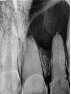
A 21 years old female patient reported to the Department of Conservative Dentistry and Endodontics with a chief complaint of pus discharge in the right anterior maxillary region since last 4 months. Medical history was noncontributory and clinical examination revealed a sinus tract in the anterior region with discolored maxillary right central incisor. Radiographic examination revealed well defined radiolucency involving the coronally discolored maxillary right central incisor and lateral incisor. Both the teeth presented a negative response to pulp testing. The treatment plan included root canal therapy followed by surgical management of the periapical lesion which included enucleation and apicectomy in relation to 11 and 12.
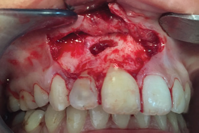
The endodontic treatment procedures were conducted. After teeth isolation the access cavities were prepared followed by shaping and cleaning. The teeth were then obturated using cold lateral condensation. Raising a trapezoidal flap, a periapical surgery was performed and periapical pathosis was enucleated. Maxillary right central and lateral incisors were apically resected 3 mm from the apex and retrograde cavity was prepared using ultrasonic tip. MTA was placed as retrograde restorative material of 3 mm thickness. The placement of the retrograde filling material was confirmed using a radiograph and the sutures were placed.
The patient was recalled after a week for suture removal. The patient was kept under observation for the purpose of postoperative evaluation of healing of the periapical intervention in intervals of 1, 3, 6, 12, and 18 months.
CASE 1

Pre Operative
Radiograph Of Obturated Canals

Bleeding Points Marked

Flap Raised
Mta Placed
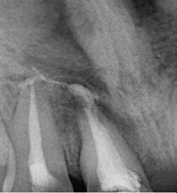
After Root End Resection
CASE REPORT 2
A 55 years old female patient reported to the Department of Conservative Dentistry and Endodontics with a chief complaint of pain in the front tooth region since last 5-6 months. No significant medical history was given. Radiographic examination revealed well defined radiolucency involving the maxillary right central incisor and lateral incisor. Both the teeth presented a negative response to pulp testing. The treatment plan included root canal therapy followed by surgical management of the periapical lesion which included enucleation and apicectomy in relation to 11 and 12.
The endodontic treatment procedures were conducted. After teeth isolation the access cavities were prepared followed by shaping and cleaning. The teeth were then obturated. Raising ochsenbein luebke flap, a periapical surgery was performed and periapical pathosis was enucleated. Maxillary right central and lateral incisors were apically resected 3 mm from the apex and retrograde cavity was prepared using ultrasonic tip. MTA was placed as retrograde restorative material. The placement of the retrograde filling material was confirmed using a radiograph and the sutures were placed.
The patient was recalled after a week for suture removal. The patient was kept under observation.
CASE 2

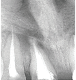
Pre Operative Pre- Operative Radiograph

Working Length Radiograph Radiograph Of Obturated Canals
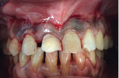
Bleeding Points Marked
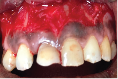
Flap Raised

Mta Placed
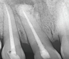
After Root End Resection
DISCUSSION
The success rates of endodontic treatment were found to decrease by 14% for every 1 mm increase in the diameter of the lesion before treatment. The negative influence of large periapical lesions is due to the presence of greater diversity of bacteria, consequently, more resistant to the treatment. This type of chronic injury, characterized by long duration, allows the micro organisms to penetrate deeper into dentinal tubules. This conta mination may even extend to the level of the apical cementum causing failures in chemical and mechanical decon tamination procedures. In addition, large lesions may also cause cystic transformation or extra-radicular infections, which render nonsurgical root canal treatment ineffective.6
Thus surgical treatment was the choice of treatment in this clinical case of the persistence of periapical lesion. Apical surgery provides an opportunity to remove the periapical lesion and the apical portion of root that has the highest anatomical variations of the root canal. Simultaneously allows a retroprepare and apical retrofilling, which aims to seal the canal system, preventing microorganisms that might still remain within the canal system, to reach the periapical tissues.7
The option for the trapezoid incision on the papilla (Neuman & Novak) is justified because of the large lesion and a triangular incision could make difficult to access and remove the lesion. A trapezoid incision on the papilla has the following advantages: excellent access to surgical field, visualization of all root length, and it is very difficult that this incision type is over the surgical cavity. Flap divulsion was initiated at the junction of the horizontal and vertical incisions so that no damage was caused to the tissue and to enable the correct flap positioning during the suture procedure.8
The use of MTA as retrofilling material is justified because it has excellent physical properties is biocompatible with the periapical tissues and has the potential of conducting and inducing cementoblasts and osteoblasts, and the ability to release the main cationic components and trigger the surface of precipitates chemically and structurally similar to hydroxyapatite. The ability of MTA to induce the cellular response is due to the phase of calcium phosphate, which can cause a change in cell behavior, simulating the adhesion of osteoblasts to this material. In addition, the MTA is non mutagenic or non-neurotoxic and produces no side effect on microcirculation.4,5
The drawbacks of MTA are related to difficulty in controlling the length of the filling, the chance of producing voids and the absence of solvent material for removal. Besides the presence of difficult handling, long setting time and high material cost.
Placing a good quality coronal restoration after root filling is considered the final step for completion of root canal treatment, because it prevents contamination of the root canal.
Concerning to post-operative progress, Howe9 stated that, generally, the patient should be followed-up at the first week after surgery and a periapical radiograph should be taken to evidence the periapical condition of the tooth at the immediate follow-up appointment. Radiographs at every three, six, 12 and 24 post-surgical months are important to determine the amount and speed of bone regeneration. In this present clinical case, the follow-up period was of 120 days (four months), at where the decreasing of the radiolucent area could be noted, evidenced the bone repair process.
CONCLUSION
Based on both the literature and the clinical case presented, it could be concluded that the endodontic surgery when well indicated and performed has a very favorable prognosis, because it eliminates the infection inside root canal, enabling root canal obturation, which consequently eliminates the infection in the periapical area, allowing bone neoformation.
REFERENCES
1. Maddalone M, Gagliani M. Periapical endodontic surgery: A 3-year follow-up study. Int Endod J 2003;36:193-8.
2. Sen BH, Piskin B, Demirci T. Observation of bacteria and fungi in infected root canals and dentinal tubules by SEM. Endod Dent Traumatol 1995;11:6-9.
3. Mortensen H, Winther JE, Birn H. Periapical granulomas and cysts. An investigation of 1,600 cases. Scand J Dent Res 1970;78:241-50.
4. Torabinejad M, Parirokh M. Mineral Trioxide Aggregate: a comprehensive literature review - Part II: leakage and biocompatibility investigations. J Endod. 2010;36(2):190-202.
5. Parirokh M, Torabinejad M. Mineral Trioxide Aggregate: a comprehensive literature review - Part III: clinical applications, drawbacks, and mechanism of action. J Endod. 2010;36(3):400-13.
6. Favieri A, Campos LC, Burity VH, Cecilia MS, Abad EC. Use of biomaterials in periradicular surgery: a case report. J Endod.2008;34(4):490-4.
7. Oliveira MAVC, Soares J, Azevedo KCM, Bifi JCG, Quirino LC, Faria RA. Apical surgery in complement to the endodontic treatment: Case report. Dental Press Endod. 2011 July-Sept;1(2):70-4.
8. Orosco et al. Endodontic surgery with simultaneous root canal filling: case report. RSBO 2014 Oct-Dec;11(4):411-413.
9. Howe GL. Cirurgia oral menor. 3 ed. São Paulo: Santos; 1990.
Disclaimer from Guident
The views and opinions expressed in articles published in Guident are solely those of the authors and do not necessarily reflect the views of the editors, the editorial board or the publisher, Ivory India. Guident does not endorse any product, technique or treatment described. Clinical content is intended for qualified dental and medical professionals and is not a substitute for individual clinical judgement. Guident, its editors and the publisher accept no liability for any consequence arising from the use of information published in the journal.
How to cite this article
Dr. Annil Dhingra, Dr. Ishita Agarwal, Dr. Shalini Sharma. Surgical Management of Large Periradicular Lesions. Guident. 2019 May;12(6). Available from: https://guident.in/articles/endodontics/surgical-management-of-large-periradicular-lesions.html