Minimum Invasive Procedure for Mandibular Impacted Third Molar Tooth ( Mipmit) - a Study to Prevent Incision and Soft Tissue Handling
Pune, Maharashtra
Abstract
Management of impacted mandibular third molars with surgical removal is very common procedure in day today practice. In which Wards or Modified Wards incisions are commonly performed. In some cases Envelope flap and other such types of flaps are commonly performed. Incision, reflection of flap, osteotomy, suturing is normal protocol which gives more tissue handling, more trauma to tissue & discomfort to patient. It also causes swelling and pain. This surgical procedure is needed in deeply seated impacted mandibular third molar tooth. But we observed in some cases of impaction, which are not deeply seated and there half or more than half crown is clinically visible in the oral cavity, we can modify the technique and we can remove this third molar with minimum invasive conservative approach. From this observation, we modified the technique for surgical removal of partially erupted third molar and inset to avoid incision and bone cutting. This paper suggests management of partially erupted mandibular third molar tooth with minimum invasive technique or Conservative approach .We observed, minimum invasive procedures causing less post-operative complications and it definitely reduces the time to perform the procedure. The prevention of incision with conservative approach is discussed in thirty patients. The purpose of this article is to present the cases that led to this observation, and present the technique modification in design and execution that we have used.[1-5]
INTRODUCTION
Mostly mesioangular & horizontal impacted tooth removal with normal protocol, more tissue handling post-operative swelling & pain is more. There is also discomfort to the patient during swallowing. To minimize these complications, author has suggested conservative and minimum invasive protocol.7-10
The individuals demand for conservative approach and stich less surgeries is increasing day by day. Through this paper author suggests conservative technique for management of mandibular third molar impacted tooth .This paper describes a new approach for removal partially impacted molar without surgical intervention & minimum trauma to tissue. This conservative approach gives less instrumentation and reduces time to perform procedure and causes minimum post-operative complications.
PATIENTS &METHOD
Thirty patients including vertical, mesioangular, horizontal, distoangular impacted mandibular third molar teeth were seen in author’s department from December 2017 to march 2018.All patients were giving history of pain in third molar region.6 Impacted tooth were confirmed by IOPA & OPG. All underwent surgical removal in aseptic condition with routine protocol of incision, bone cutting, suturing after necessary investigations.
INITIAL PROTOCOL
For this, mandibular third molar which are partially erupted into the oral cavity are selected. After administration of Local anesthesia, Incision was given with 15 number blade. With the help of periosteal elevator full thickness mucoperiosteal flap was raised .With the help of bone cutting bur, guttering of bone was performed with normal saline irrigation. Tooth was elevated with straight elevator and tooth was removed. Simple interrupted sutures was given with 3-0 mersilk. Iodinated gauze was placed and post-operative instructions were given and recalled patient after 3rd and 7th day.11
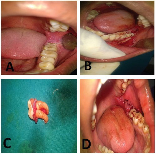
Patient treated for impacted mandibular third molar with surgical approach. A pre-operative clinical photograph, B incision and flap raised, C tooth removed, D sutures given.
NEW PROTOCOL
“MINIMUM INVASIVE PROCEDURE FOR MANDIBULAR IMPACTED THIRD MOLAR TOOTH ( MIPMIT)-A STUDY TO PREVENT INCISION AND SOFT TISSUE HANDLING.”
Thirty patients underwent mandibular third molar impacted tooth removal with following new protocol with conservative approach. For this protocol the tooth which are partially visible into the oral cavity are selected.
Local anesthesia with adrenaline was injected, with the help of straight diamond cylindrical bur with air rotor hand piece the cusp of impacted 3rd molar which is in contact with adjacent 2nd molar is trimmed without damaging 2nd molar & contact point is broken in between 2nd& 3rd molar. With help of moon’s probe reflection of gingiva was done. If there is no purchase point for elevator then with help of straight or round bone cutting bur purchase point was created with the help of straight hand piece attached to micro motor .With the help of straight elevator 3rd molar is elevated. Elevation is in such a manner that crown is pulled buccally & then upward so that roots of tooth moved lingually & then downward, due to thin lingual cortical plate, this movement can happen easily and it gives space and path of removal for 3rd molar without hitting 2nd molar. We observed that Post-operative complications like swelling, pain and trismus were reduced as compare to routine surgical approach.16
CASE 1
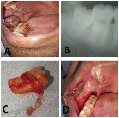
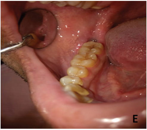
Minimum invasive conservative technique for removal of mandibular third molar tooth. A pre-operative photograph, B pre-operative x-ray, C tooth removed with conservative approach, D post-operative minimum trauma without incission, E post-opperative 7th day healing.
CASE 2
Minimum invasive conservative technique for removal of mandibular third molar tooth. A pre-operative photograph, B pre-operative x-ray, C tooth removed with conservative approach, D post-operative minimum trauma without incission.

CASE 3
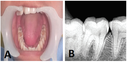
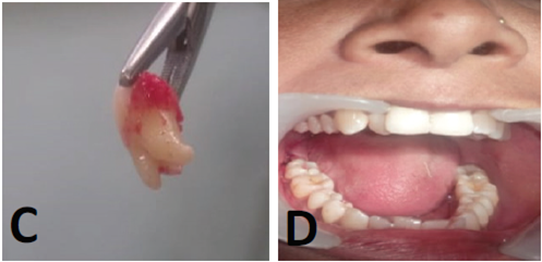
Minimum invasive conservative technique for removal of mandibular third molar tooth. A pre-operative photograph, B pre-operative x-ray, C tooth removed with conservative approach, D post-operative minimum trauma without incission.
RESULTS
The present study included 60 clinically and rediographycally confirmed partially impacted third molar tooth. IOPA or OPG taken for every cases.12-15.30 cases were operated with normal protocol of incision, bone cutting, tooth removal and suturing. Another 30 cases were operated with author’s minimum invasive conservative approach. Tissue injury, bone loss, post-opperative pain, swelling ,trismus observed less in modified technique as compare to routine surgical approach. Even the post-operative wound was very small like simple extraction and most of the cases, there was no need to suture it. Healing was faster as compare to routine surgical technique.18-19
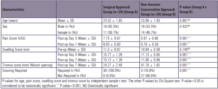
Table 1 Inter-group comparison of study characteristics.
Distribution of Mean Age
The mean ± SD of age of cases studied in Group A and Group B was 23.52 ± 23.60 years and 23.60 ± 1.83 years respectively. The minimum – maximum age range in Group A and Group B was 20-28 years and 21-28 years respectively.
The distribution of mean age of cases studied did not differ significantly between two study groups (P-value>0.05).
Distribution of Sex
Of 30 cases studied in Group A, 19 (63.3%) were male and 11 (36.7%) were female. Of 30 cases studied in Group B, 16 (53.3%) were male and 14 (46.7%) were female.
The sex distribution of cases studied did not differ significantly between two study groups (P-value>0.05).
Distribution of Mean Pain Score (VAS – visual analogue scale)
The mean ± SD of pain score (VAS) at post-op day 3 among the cases studied in Group A and Group B was 7.73 ± 0.87 and 0.87 ± 0.86 respectively.
The distribution of mean pain score (VAS) at post-op day 3 is significantly higher in Group A compared to Group B (P-value<0.001).
The mean ± SD of pain score (VAS) at post-op day 7 among the cases studied in Group A and Group B was 5.44 ± 0.81 and 0.82 ± 0.52 respectively.
The distribution of mean pain score (VAS) at post-op day 7 is significantly higher in Group A compared to Group B (P-value<0.001).
Distribution of Mean Swelling Score
The mean ± SD of pre-op swelling score among the cases studied in Group A and Group B was 10.94 ± 1.13 cm and 10.43 ± 1.09 cm respectively.
The distribution of mean pre-op swelling score did not differ significantly between two study groups (P-value>0.05).
The mean ± SD of swelling score at post-op day 3 among the cases studied in Group A and Group B was 16.39 ± 1.49 cm and 11.03 ± 1.12 cm respectively.
The distribution of mean swelling score at post-op day 3 is significantly higher in Group A compared to Group B (P-value<0.001).
The mean ± SD of swelling score at post-op day 7 among the cases studied in Group A and Group B was 15.61 ± 1.33 cm and 11.00 ± 1.03 cm respectively.
The distribution of mean swelling score at post-op day 7 is significantly higher in Group A compared to Group B (P-value<0.001).
Distribution of Mean Trismus Score (Mouth opening score)
The mean ± SD of Trismus score at post-op day 7 among the cases studied in Group A and Group B was 24.27 ± 2.48 mm and 41.70 ± 1.62 mm respectively.
The distribution of mean Trismus score at post-op day 7 is significantly higher in Group B compared to Group A (P-value<0.001).
Distribution of Requirement of Suturing
Of 30 cases studied in Group A, 30 (100.0%) required suturing. Of 30 cases studied in Group B, 3 (10.0%) required suturing and 27 (90.0%) did not require suturing.
The distribution of requirement of suturing is significantly higher in Group A compared to Group B (P-value<0.001).
DISCUSSION
The individuals demand for conservative approach and stichless surgeries is increasing day by day. The intraoral mucosa which is incised in normal surgical protocol belongs to patient. The bone which used to cut during surgery belongs to patient. So surgeon have to think to preserve the mucosal tissue and bone at his best possible. Author observed that in his minimum invasive approach we are trimming the tooth at contact point which has to be removed which is waste product for patient. Only modification in elevating the tooth is needed in this approach. We have to elevate tooth bucally and upward to create path of removal without touching adjacent second molar.It happens easily as crown moves bucally, root portion moves lingually easily due to less thickness of lingual cortical plate.16
CONCLUSION
Impacted tooth which is not visible in oral cavity clinically then normal routine surgical protocol is needed to remove the impacted mandibular third molar. But we can preserve tissue and bone with this minimum invasive modified technique in some cases of partial erupted mandibular third molar tooth which is indicated for removal.17 It is observed that MIPMIT technique gives less post-operative pain, swelling and trismus. Healing is also faster than routine surgical technique. Mostly post-operative wound was like normal tooth extraction so there was no need for sutures in most of the cases. The time taken for procedure was very less as compare to the surgical protocol. Author suggests that this is revolutionary technique for the society.
Ethical Approval
This study was approved by the ethics committee of the Institution. All procedures performed in the study were in accordance with the ethical standards of the institutional ethics committee.
Informed consent
Informed written consent was obtained from the patient included in the study.
REFERENCES
- .ouloux G, Steed M, Perciaccante V (2007) Complications of Third MolarSurgery. Oral Maxillofacial SurgClin N Am 19: 117-128.
- .marillas E, Toranzo J, Martínez R, Loyola M, Hidalgo J, et al. (2010) Use ofTherapeutic Laser After Surgical Removal of Impacted Lower Third Molars. JOral MaxillofacSurg 68: 319-324.
- Farish S, Bouloux G (2000) General Technique of Third Molar Removal. OralMaxillofacial SurgClin N Am 19: 23-43.
- Susarla S, Dodson T (2007) Preoperative Computed Tomography Imaging inthe Management of Impacted Mandibular Third Molars. J Oral MaxillofacSurg 65: 83-88.
- Graziani F, D’Aiuto F, Arduino P, Tonelli M, Gabriele M (2006) Perioperativedexamethasone reduces post-surgical sequelae of wisdom tooth removal. Asplit mouth randomized double-masked clinical trial. IntJOralMaxillofacSurg35: 241-246.
- Adeyemo WL, James O, Ogunlewe MO, Ladeinde AL, Taiwo OA, et al. (2008)Indications for extraction of third molars: a review of 1763 cases. Niger PostgradMed J 15: 42-46.
- Susarla S, Blaese Br, Magalnick D (2003) Third molar surgery and associatedcomplications. Oral Maxillofacial SurgClin N Am 15: 177-186.
- Pozos A, Martínez R, Aguirre P, Pérez J (2007) Tramadol Administered in aCombination of Routes for Reducing Pain After Removal of an ImpactedMandibular Third Molar. J Oral MaxillofacSurg 65: 1633-1639.
- Kirmeier R, Truschnegg A, Payer M, Acham S, Schulz K, et al. (2007) Evaluation of a muscle relaxant on sequelae of third molarsurgery: a pilot study. Oral SurgOral Med Oral Pathol Oral RadiolEndod 104: e8-e14.
- Blondeau F, Daniel N (2007) Extraction of Impacted Mandibular Third Molars: Postoperative Complications and Their Risk Factors. JCDA 73: 325
- Abu-El Naaj I, Braun R, Leiser Y, Peled M (2010) Surgical Approach toImpacted Mandibular Third Molars-Operative Classification. J Oral MaxillofacSurg 68: 628-633.
- Guillaume B, Lacoste JP, Gaborit N, Brossard G, Cruard A, et al. (2006)Microcomputed tomography used in the analysis of the morphology of rootcanals in extracted wisdom teeth. British Journal of Oral and Maxillofacial Surgery 44: 240-244.
- Wagner KW, Otten JE, Schoen R, Schmelzeisen R (2005) Pathologicalmandibular fractures following third molar removal. Int J Oral MaxillofacSurg34: 722–726.
- Lübbers HT, Matthews F, Damerau G, Kruse AL, Obwegeser JA, et al.(2011) Anatomy of impacted lower third molars evaluated by computerizedtomography: is there an indication for 3-dimensional imaging? Oral Surg Oral Med Oral Pathol Oral RadiolEndod 111: 547-550.
- Kunkel M, Doz P, Morbach T, Kleis W, Wagner W (2006) Third molar complications requiring hospitalization. Oral Surg Oral Med Oral Pathol OralRadiolEndod 102: 300-306.
- Kim HR, Choi BH, Engelke W, Serrano D, Xuan F, et al. (2011) A comparativestudy on the extractions of partially impacted mandibular third molars with orwithout a buccal flap: a prospective study. J Oral MaxillofacSurg 69: 966-970.
- White RP Jr, Madianos PN, Offenbacher S, Phillips C, Blakey GH, et al. (2002)Microbial complexes detected in the second/third molar region in patients withasymptomatic third molars. J Oral MaxillofacSurg 60: 1234-1240.
- Flynn TR, Shanti RM, Levi MH, Adamo AK, Kraut RA, et al. (2006) Severeodontogenic infections, part 1: prospective report. J Oral MaxillofacSurg 64:1093-1103.
- MarcianiRD: Third Molar Removal: An Overview of Indications, Imaging, Evaluation,and Assessment of Risk: Oral Maxillofacial SurgClin N Am 19 (2007) 1–13
- Textbook and color atlas of tooth impactions: Anderson, Peterson, LaskinOMF Clinics of North America 19(2007), 59-68
- Impacted third molar-Dimitraulis
- Textbook of oral surgery: Archer
Disclaimer from Guident
The views and opinions expressed in articles published in Guident are solely those of the authors and do not necessarily reflect the views of the editors, the editorial board or the publisher, Ivory India. Guident does not endorse any product, technique or treatment described. Clinical content is intended for qualified dental and medical professionals and is not a substitute for individual clinical judgement. Guident, its editors and the publisher accept no liability for any consequence arising from the use of information published in the journal.
How to cite this article
Dr. Nikam Vishwaraj Hanumant. Minimum Invasive Procedure for Mandibular Impacted Third Molar Tooth ( Mipmit) - a Study to Prevent Incision and Soft Tissue Handling. Guident. 2019 May;12(6). Available from: https://guident.in/articles/research/minimum-invasive-procedure-for-mandibular-impacted-third-molar-tooth-mipmit-a-study-to-prevent-incision-and.html