Magnification in Dentistry-Introduction to Magnification
of upskill Dental Academy, Mumbai, India
From time immemorial man has been in continuous pursuit to see things far smaller than can be seen with normal vision. During the 1st century AD, glass had been invented and the Romans were looking through the glass and experimenting with it. They discovered that if they held one glass which is thick in the centre and thin at the margins over an object, the object would appear much larger.
It was Antony Van Leeuwenhoek (1632-1723), a Dutch draper and scientist, and one of the pioneers of microscopy who in the late 17th century became the first man to make and use a real microscope. This opened the world of magnification.
The Merriam-Webster defines magnification as the apparent enlargement of an object by an optical instrument-called also as power.
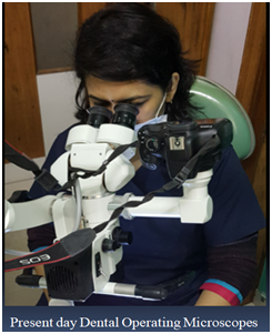
Magnification has been introduced to dentistry through dental loupes and dental operating microscopes.
However its acceptance and application is taking time. It becomes essential for us to understand what is magnification and what is its relevance in routine dentistry.
HISTORY OF DENTAL OPERATING MICROSCOPES
Operating microscopes have been used for decades in many medical disciplines : ophthalmology, otorhinolaryngology, neurosurgery, vascular surgery ,however its introduction to dentistry has been many years later.1
The words of Prof. Syngcuk Kim “you can only treat what you can see!” set the thought process.
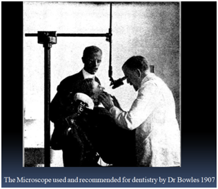
In 1978 Dr. Apothekar and Dr Jako brought the concept of extreme magnification, in the form of an operating microscope, into dentistry. They postulated that the tremendous improvements in visual acuity, made possible through the use of the operating microscope, would be beneficial to the discipline of Endodontics.
The first commercially available Dental Operating microscope named Dentiscope was introduced in 1981.its market failure was more a function of its poor ergonomic design than its good optical properties.
In the late 1980s, San Deigo endodontist Dr. Gary Carr concluded that incredible magnification and illumination made possible with the microscope could be beneficial to endodontists and started promoting its usage as a crucial piece of armamentarium used in improvement of outcomes of endodontic apical surgeries. Dr Howard Seldon (1986) was the first endodontist to publish an article on the use of dental operating microscopes in Endodontics where he discussed its use in conventional treatment of a tooth, not in surgical Endodontics.
In 1995, the American Association of Endodontists (AAE) formally recommended to the Commision on Dental Accredition (CODA) of the American Dental association (ADA) that microscopy training be included in the new accredition standards for the Advanced speciality Education programmes in Endodontics. In January 1997, the new standards, making microscopy training mandatory, became effective.
In 1999 Dr Gary Carr, introduced the dental operating microscope that had Gallilean optics and that was ergonomically configured for dentistry, with several advantages that allowed the easy use of scope for nearly all endodontic and restorative procedures. 2
UNDERSTANDING MAGNIFICATION
THE HUMAN EYE
The human eye is easily one of the most complex organs of the body .If we consider its working in detail...Light enters the eye through the cornea, the clear front surface of the eye. The iris works much like a diaphragm of a camera-controlling how much light reaches the back of the eye. This is done by automatically adjusting the size of the pupil. The eye’s crystalline lens is present exactly behind the pupil. Focussed by the cornea and the crystalline lens, the light makes its way to the retina. It is at the retina the process of image formation takes place. To conclude the eye is an optical system at work.
CAN THE HUMAN EYE MAGNIFY ?
We have to be familiar with the term near point of the eye. The near point of the eye is the point nearest the eye at which an object can be placed and still have a sharp image produced on the retina. For the normal eye, the nearest point is located 25cm from the eye. It also the point at which the object appears largest to the eye.
LIMITATIONS OF THE EYE
For a clinician to view a tooth enlarged at maximum resolution naturally he/she will have to be at a distance of 25cm from it. This is impractical in a clinical set up. Secondly although the clinicians have routinely strived to create bacteria free seals, the resolving power of unaided human eye is only 0.2mm. in other words most people who view two points closer than 0.2mm will see only one point. Clearly dentists are at a severe disadvantage in their attempts to replace natural tooth structure with artificial materials that do not leak, in view of virtually invisible microbiologic threats to restoration integrity.1
THE SOLUTION
The scenario can be reproduced and a better magnification can be obtained by placing an optical lens system between the eye and the object (tooth). This is accomplished without compromising on the operator posture .This emphasises the role of magnification in a dental set up.
The prime purpose of a clinician to invest in magnification especially in a microscope is usually for better precision .however along with precision they are benefitted with superior ergonomics, excellent patient communication tool and high intensity illumination .
The microscope can completely change the quality of routine dentistry.
BiBLOGRAPHY
- The use of the Operating Microscope in Endodontics. Prof Gary B. Carr and Arnaldo Castelluci.
- Dental Operating Microscope in Endodontics –A Review. Prof.(Dr)Utpal Kumar Das, Dr Subhasis Das.
Disclaimer from Guident
The views and opinions expressed in articles published in Guident are solely those of the authors and do not necessarily reflect the views of the editors, the editorial board or the publisher, Ivory India. Guident does not endorse any product, technique or treatment described. Clinical content is intended for qualified dental and medical professionals and is not a substitute for individual clinical judgement. Guident, its editors and the publisher accept no liability for any consequence arising from the use of information published in the journal.
How to cite this article
Dr.Pavana Kamath Director. Magnification in Dentistry-Introduction to Magnification. Guident. 2018 May;11(6). Available from: https://guident.in/articles/endodontics/magnification-in-dentistry-introduction-to-magnification.html