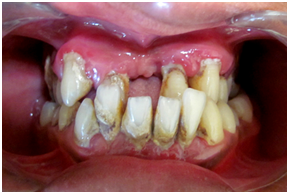
Platelet -Rich Fibrin in Regeneration of Intrabony Defects
The Oxford Dental College, Banglore, India
ABSTRACT
Although periodontitis is an infectious disease of periodontal tissues, changes that occur in the bone are crucial, because destruction of bone is responsible for tooth loss. Although horizontal bone loss is most common, vertical bone loss is more amenable to regenerative periodontal therapy. Recently, importance has been given to use of platelet-rich fibrin (PRF) for predictably obtaining periodontal regeneration. PRF is a concentrated suspension of growth factors found in platelets. These growth factors moderate the wound healing and promote tissue regeneration. This article includes case report of a patient where an infrabony defect was treated using PRF. This case report shows the clinical and radiographical outcome obtained by use of PRF in intrabony defect. Key words: Intra bony defect, PRF, Regeneration
INTRODUCTION:
The biologic goal of periodontal regeneration is restoration of the periodontium to its original form and function. Periodontal repair is "healing of the periodontium by tissue that does not fully restore the original function, anatomic and morphologic architecture." Following conventional surgical approaches, such as open flap debridement, repair of lost periodontal attachment occurs primarily through formation of a long junctional epithelium between the gingival flap and the previously diseased root surface.1 Regeneration has been defined as "the reproduction or reconstitution of a lost or injured part to restore the architecture and function of the periodontium."2 The aim of regenerative periodontal therapy is to restore the structure and function of the periodontium. Periodontal regeneration requires an orchestrated sequence of biologic events such as cell migration, adherence, growth and differentiation, to have the potential to increase the success and predictability of periodontal regenerative procedures.3
According to a position paper from the American Academy of Periodontology, periodontal regenerative procedures include soft tissue grafts, bone replacement grafts, root biomodifications, guided tissue regeneration (GTR), and combinations thereof for osseous, furcation, and recession defects.4
Platelet-rich fibrin (PRF) was first described by Choukroun et al. in 2004 in France. It has been referred to as a second generation platelet concentrate5 because the natural concentrate is produced without any anticoagulants or gelifying agents. First biochemical analysis of the PRF composition indicated that this biomaterial consists of an intimate assembly of cytokines, glycanic chains, and structural glycoproteins enmeshed within a slowly polymerized fibrin network. This biologic activity of fibrin molecule is by slow polymerization mode, which confers the PRF membrane a favorable physiological architecture to support the healing process.2
A recent innovation in the dentistry is the preparation and the use of PRF, a concentrated suspension of the growth factors found in platelets. These growth factors are involved in wound healing and are postulated as promoters of tissue regeneration. Platelet concentrate contains platelet-derived growth factor (PDGF), transforming growth factor (TGF), vascular endothelial growth factor (VEGF), etc. growth factors that modulate and up-regulate growth factor function.6
Here, we present a 6-month follow-up report of an intrabony defect treated with an autologous PRF by assessing clinical and radiological parameters.
CASE REPORT:
Patient reported to the Department of Periodontics, The oxford dental college, Bangalore with the complaint of food lodgement and pain in the gingiva. There was no any relevant medical history, no history of dental trauma, orthodontic treatment, and no injurious habit was reported.
A 32-year-old male complained of food lodgement and pain in the lower right mandibular molar
region. On intraoral examination, there was bleeding on probing present no swelling and no pus exudation were noticed. The probing pocket depth (PD) on the mesiobuccal aspect of tooth #46 was 15 mm, grade II furcation involvement and grade I mobility was detected. A periapical radiograph was taken. Radiographs were standardized by using paralleling technique and holder, which revealed presence of interproximal angular bone loss. (Fig 1) The following treatment plan was made: -
- Non-surgical periodontal therapy by means of conventional scaling and root planning using ultrasonic instrument and curettes. -
- Patient was recalled after every week and re-examination was done for 4 weeks. -
- Surgical periodontal therapy was planned since there was persistant periodontal pocket of about 14mm was present with grade II furcation involvement.
A complete blood investigation was done which included platelet count, hemoglobin, bleeding time, and clotting time were assessed and found to be within normal limits.
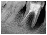
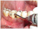
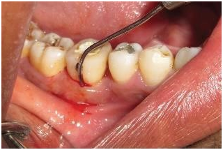
Fig 1: pre operative IOPA (a), probing depth (14mm) (b) and furcation involvement(c)(Grade II)
PRF PREPARATION
The PRF was prepared in accordance with the protocol developed by Choukroun et al.7 just prior to surgery, intravenous blood from antecubital vein was collected in the 10 ml of sterile tube without an anticoagulant and centrifuged immediately. Blood was centrifuged using a tabletop centrifuging machine for 12 min at 2,700 rpm (Fig 2). PRF was easily separated from RBCs using a sterile tweezer just after removal of PPP
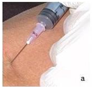
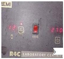
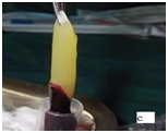
Fig.2: Preparation of platelet rich fibrin memebrane.
(a) Blood withdrawn from antecubital vein; (b) centrifuged at 2700rpmfor 12 minutes; (c) the platelet rich fibrin (PRF) obtained.
SURGICAL PROCEDURE
Intraoral antisepsis were performed using 0.2% chlorhexidine digluconate rinse. Following administration of local anesthesia, sulcular incisions were made, and mucoperiosteal flap were reflected. Meticulous defect debridement and root planning was carried out with the help of area specific curettes. The direct examination after debridement, confirmed the presence of one walled bony defect. PRF was filled into the infrabony defect. The mucoperiosteal flap were repositioned and secured in place using 3-0 non-absorbable black silk surgical suture. The simple interrupted sutures were placed (Fig.3).
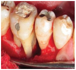
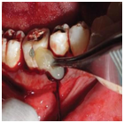


Fig 3 : (a) Mucoperiosteal flap raised (Infrabony defect seen irt 45-46); (b) PRF placed ; (c) Interrupted sutures placed; (d) Periodontal pack placed.
POST OPERATIVE CARE:
Antibiotics and analgesics (amoxicillin 500 mg three times a day for 5 days and Diclofinac + Paracetamol two times a day for 5 days) were prescribed along with 0.2% chlorhexidine mouth wash twice daily, for 1week. Periodontal pack and sutures were removed after 1 week postoperatively. Surgical wound were cleaned with iodine and patients were instructed for gentle brushing with soft toothbrush. Patients were instructed for oral hygiene maintenance and reexamined at 1 week, 2 month and 3 months, which showed reduction in mobility and pocket depth, with significant bone fill. (Fig 4)

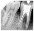
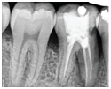
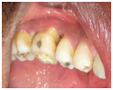
Fig4: one week (a); two month (b); three month post-op IOPA (c); (d) post-op clinical view
DISCUSSION
The ideal goal for periodontal therapy is the reconstitution of bone and connective tissue attachment that has been destroyed by the disease process.8 The complete regeneration of the periodontium after periodontal treatment modalities has been difficult to achieve because of differences in the healing abilities among periodontal tissues. During the last 12 years, the field of periodontics has made great strides in developing techniques to regenerate the lost tissues. Consequently, dental research and industry have been increasingly concerned over the past two decades with biologically inert, synthetic, and autologous materials for implantation into periodontal infrabony defect. The present case report evaluated the clinical efficacy of PRF in the treatment of infrabony defects in patients with chronic periodontitis and showed a significant improvement in clinical and radiographic parameters. Platelet concentrates can be used as regenerative tools as platelets possess growth factors and cytokines that can be harnessed for bone regeneration and soft-tissue maturation. PRF does not dissolve quickly after application but the strong fibrin matrix is remodeled slowly in a manner similar to a natural blood clot. PRF membrane has a very significant sustained release of key growth factors for at least one week and upto 28 days according to research. Apart from platelets, leucocytes and fibrin in platelet concentrates also have significant roles. Leukocytes have an anti-infectious action and aids in immune regulation. They produce large amounts of VEGF to promote angiogenesis and serve as a biological healing matrix by supporting cell migration and cytokine release. Hence, PRF can be employed as a potential adjunct in treatment of intrabony periodontal defects.9 Anuj Sharma et al. (2011)10 and Thorat et al. (2011)11 found that there was greater pocket depth reduction, clincal attachment level (CAL) gain, and bone fill at the sites treated with PRF with
conventional open-flap debridement compared to conventional open-flap debridement alone. Simonpieri et al. (2009)12 & Patel et al, (2017)9 described the four advantages of the use of PRF during bone grafting. First, fibrin clot plays an important mechanical role in maintaining and protecting the graft and PRF fragments serve as biological connectors between bone particles. Second, fibrin network facilitates cellular migration, vascularization, and survival of the graft. Third, the growth factors (PDGF, TGF-β, IGF-1) are gradually released as the fibrin matrix is resorbed, thus creating a perpetual process of healing. Lastly, the presence of leukocytes and cytokines in the fibrin network can play a important role in the self-regulation of inflammatory and infectious phenomena within the grafted material. Further, PRF in combination with other bone substitutes such as bovine porous bone mineral and hydroxyapatite or alone resulted in improvement in clinical and radiographic parameters in the treatment of human three wall intrabony defects.13, 14 There are wide range of regenerative procedures that have been developed over the years. It is important to recognize the benefits of each process and use them in indicated situations. The patients could be benefited in such situations, by maximum possible regeneration, ideally reaching physiological levels.
CONCLUSION
From the presented case, it can be concluded that PRF is efficacious clinically and radiographically in the periodontal infrabony defect. PRF is an autologous preparation and found to be clinically and economically effective than any other available regenerative material. However, long-term, multicenter randomized, controlled clinical trials will be required.
REFERENCES:
- Reynolds MA, Aichelmann-Reidy ME, Branch-Mays GL. Regeneration of Periodontal Tissue: Bone replacement grafts. Dent Clin North Am 2010;54:55-71.
- Bajaj P, Rao NS, Agarwal E, Pradeep AR. Treatment of intrabony defect with platelet rich fibrin: A case report. Arch Oral Sci Res 2011;1:90-4.
- Pradeep AR, Shetty SK, Garg G, Pai S. Clinical effectiveness of autologous platelet-rich plasma and Peptide-enhanced bone graft in the treatment of intrabony defects. J Periodontol 2009;80:62-71.
- Greenwell H; Committee on Research, Science and Therapy. American Academy of Periodontology. Position paper: Guidelines for periodontal therapy. J Periodontol 2001;72:1624-8.
- Dohan DM, Choukroun J, Diss A, Dohan SL, Dohan AJ, Mouhyi J, et al. Platelet-rich fibrin (PRF): A second-generation platelet concentrate. Part II: Platelet-relate biologic features. Oral Surg Oral Med Oral Pathol Oral Radiol Endod 2006;101:e45-50.
- Wartiovara U, Salven P, Mikkola H, Lassila R, Kaukonen J, Joukov V, et al. Peripheral blood platelets express VEGF C and VEGF which are released during platelets activation. Thromb Haemost 1998;80:171-5.
- Choukroun J, Adda F, Schoeffler C, Vervelle A. An opportunity in perio-implantology: The PRF. Implantodontie 2000;42:55-62.
- Zander HA, Polson AM, Heijl LC. Goals of periodontal therapy. J Periodontol 1976;47:261-6.
- Patel GK, Gaekwad SS, Gujjari SK, S C VK. Platelet-Rich Fibrin in Regeneration of Intrabony Defects: A Randomized Controlled Trial. J Periodontol. 2017 Nov;88(11):1192-1199.
- Sharma A, Pradeep AR. Treatment of 3-wall intrabony defects in patients with chronic periodontitis with autologous platelet-rich fibrin: A randomized controlled clinical trial. J Periodontol 2011;82:1705-17.
Disclaimer from Guident
The views and opinions expressed in articles published in Guident are solely those of the authors and do not necessarily reflect the views of the editors, the editorial board or the publisher, Ivory India. Guident does not endorse any product, technique or treatment described. Clinical content is intended for qualified dental and medical professionals and is not a substitute for individual clinical judgement. Guident, its editors and the publisher accept no liability for any consequence arising from the use of information published in the journal.
How to cite this article
Dr. Neethu.P Dr. Anirban Chatterjee Dr. Amita Countinho Department of Periodontology. Platelet -Rich Fibrin in Regeneration of Intrabony Defects. Guident. 2018 May;11(6). Available from: https://guident.in/articles/periodontics/platelet-rich-fibrin-in-regeneration-of-intrabony-defects.html