
Dental Operating Microscope its Maintenance
Director, Upskill Dental Academy Mumbai, India
Dental operating microscope is a life time investment. A knowledge of the possible source of damage to lens with due course of time is essential to the operators. One known cause being damage caused due to fungus.
Lens fungus Lens fungus is a microbial growth on glass surfaces of optical devices. It is very common. It takes a number of forms but the typical destructive growth pattern results from microscopic spores growing in webbed, branching mycelium structures across an optical surface.
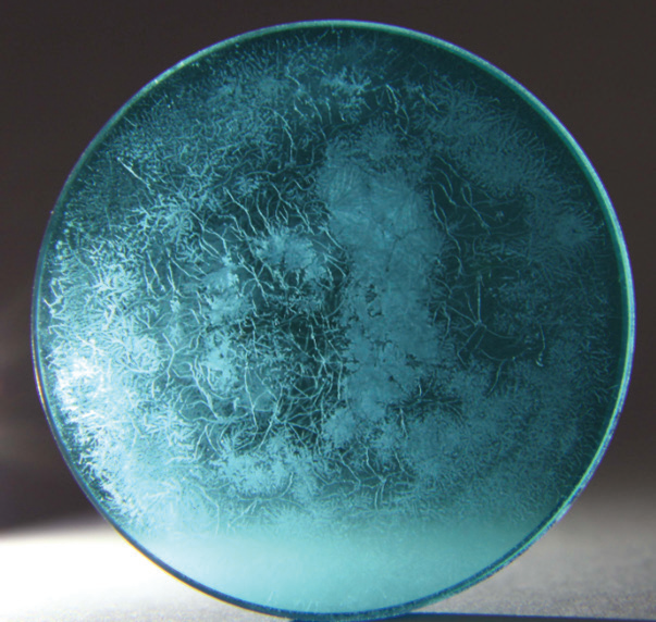
Source Internet
In some cases this can be cleaned and the lens restored. Metabolic products of the fungus etch the glass lens surface or coatings, or the growth may be on internal glass surfaces that cannot be reached. Like dust or fingerprints on a lens, obstruction and obscuration of the lens progress in degrees, from a slight loss of image contrast and resolution to complete loss.
Fungal growth on the lens
Mechanism of growth and subsequent In hot and humid climates it is common for fungus to grow on the surfaces of optical components. Airborne fungal spores settle on optical surfaces and develop into organisms that digest organic material, such as oils from fingerprints or lens coatings, producing hydrofluoric acid as a waste product. This acid in turn destroys any lens coatings and permanently etches the glass.
The dust in the surroundings consists of carbohydrate (cellulose lint from fabrics) or protein that is fine food for fungus. This dust gets pushed into the corners of a lens from cleaning, where it collects, and these marginal heaps also tend to retain humidity. Humidity above 60 percent is considered to be the threshold where fungal growth starts. All it takes is one spore to germinate on a single tiny lint fiber, which gives rise to the fibrils across the glass. A fungal spore can feed on a tiny speck of dust for many days, covering many square millimeters of area with fibrils that will find other food present. Live fungus is basically a molecular machine that is powered by the minute chemical energy available in the dust, which dust is also digested into the substance of the fungal structure. This growth mechanism converts bits of energy-rich food into less energetic but more connected and purposed structures. The structures send (mycelium fibrils) to locate and tap more energy sources, in hopes of finding a source of energy that will supply enough energy and material to launch (spores) into the air.
The growth on the lens is typically does not release spores, the resulting mycelium will degrade the lens. Besides the mechanical obstruction of the light rays the lens is designed to refract and transmit, the mycelium growth produces chemical by products which etch the glass.
Glass as a source for fungal Optical glass can have a widely varied composition. The etched surface of glass becomes a physical substrate which anchors or promotes growth. The highly detailed structure of the etching suggests that whatever metabolic product is disintegrating glass is concentrated and bound to the organism.
How to protect the optical lens /avoid fungal growth
- Lens fungus is a constant, progressive, accumulating threat. Keep the optical instrument away from warmth and humidity
- To prevent fungal growth on optical components the following precautions should be observed.
- After instruments with optics have been used and cleaned, they should be dried immediately. Turning on a fan in the room will hasten the drying process.
- Keep optical components in a dry place with a relative humidity of less than 65% and with plenty of air circulation. Air conditioners and dehumidifiers are very helpful, but must be used 24 hours a day since sudden changes in temperature and humidity promote the growth of fungus.
- In a humid environment, do not cover optics with plastic drape covers (commonly supplied by medical equipment manufacturers) since these will retain humidity. If you need to drape the equipment to protect against dust, use a cloth cover. Do not use containers made of leather, textiles or wood to store optic.
- Exposing optics to short periods of sunlight or artificial UV light may help prevent fungal growth. An operating microscope which is constantly functional has less frequent problems related to fungal growth.
- Use replaceable fungicidal pellets in cabinets where optics are stored or inside large devices such as surgical microscopes. These can be obtained from some instrument manufacturers, and have a useful life of about 3 years.
Removing fungus
Removing fungus from lenses can be difficult and may not yield suitable results since the damage is often permanent. Killing and removing the fungus and cleaning the optical surface may prolong the useful life of the instrument, if it can still provide an acceptable image.
REFERENCES
- KNOW YOUR LENS FUNGUS
Identifying and Preventing the Ruin of Camera and Instrumentation Lenses
Richard J Kinch
http://www.truetex.com
Updated: April, 2019.
- Fungus: how to prevent growth and remove it from optical components
Ismael Cordero
Community Eye Health. 2013; 26(83): 57.
Disclaimer from Guident
The views and opinions expressed in articles published in Guident are solely those of the authors and do not necessarily reflect the views of the editors, the editorial board or the publisher, Ivory India. Guident does not endorse any product, technique or treatment described. Clinical content is intended for qualified dental and medical professionals and is not a substitute for individual clinical judgement. Guident, its editors and the publisher accept no liability for any consequence arising from the use of information published in the journal.
How to cite this article
Dr. Pavana Kamath. Dental Operating Microscope its Maintenance. Guident. 2019 Nov;12(12). Available from: https://guident.in/articles/endodontics/dental-operating-microscope-its-maintenance.html


