
Conservative Management of Intracanal Separated Endodontic Instruments Using File Bypass Technique- a Case Series
Department of Conservative Dentistry and Endodontics, Seema Dental College And Hospital, Rishikesh
INTRODUCTION
The potential for instrument separation is present during every standard endodontic treatment. This complication can have an enormous impact on the outcome of the treatment, can be an obstacle for the proper cleaning, shaping and definitive obturation of the root canal system and can therefore be the reason for a future endodontic failure.1
Frequency of instrument separation: The separation rate of Nickel Titanium (NiTi) rotary instruments were reported to range between 1.3% and 10.0%, whereas separation rates of stainless steel (SS) instruments were reported to range between 0.25% and 6%.2
Instrument separation can be the result of:
- Improper use of the instrument
- Inadequate access
- Unusual and unexpected Root Canal System anatomy
- Manufacturing defects
- Rush to use rotary instruments without creating a proper straight-line access and glide path
- Higher breakage rate of rotary files is proved in cases when the root canal was not previously prepared with hand files
- Not following the rule that files have to advance slow in the root canal, until resistance is felt
- Not creating glide path to the working length using #10 and #15 files
- Overuse of endodontic instruments1
The four treatment protocols have been suggested by the literature for management of fractured instruments in root canals:
- Allowing the separated instrument to be retained in the canal and treating the remaining portion of canal.
- Bypassing the separated fragment and treating the canal.
- Retrieving the separated fragment and treating the canal.
- Surgical approach for retrieval of separated fragment followed by treatment accordingly.2
Bypassing the instrument is conservative procedure if retrieval of the instrument fails. It is the technique of going around a fractured instrument with another instrument usually a smaller one. In this way the root canal beyond the fractured instrument can be clean and the fracture instrument is incorporated in the root canal filling3 The bypassing procedure is considered more conservative and less structurally invasive because it does not require such excessive dentin removal from the root canal wall. It is a relatively simple procedure that often results in complete removal of the fragment and allows the achievement of full working length, proper preparation, irrigation and obturation of the root canal system.1
CASE 01:
A 24 year old girl reported to the department with dull pain in the right lower back region for the last one month. Radiographic examination revealed dental caries in the right lower 1st molar tooth. After elaborate history taking and clinical examination, diagnosis of dental caries with chronic irreversible pulpities w.r.t 46 was made. Root canal treatment was performed as follows: Access opening was done and working length was determined. During shaping and cleaning, a file was separated in distobuccal canal of the treated tooth. A radiograph was taken to confirm the level of separation of the instrument. The instrument was found to be separated at the apical 3rd of the distal canal. On clinical examination, there was no tenderness, mobility or swelling. However, as the broken file was remained within the canal and there was no periapical pathology, a nonsurgical file bypass technique was selected for this case. A radiograph was taken to confirm the position of separated file in canal and then bypass technique was applied as follows: With glide path the fragment was tried to loosen with # 8 file and then inserted the file slowly and carefully into the canal, and tried to negotiate past the fragment in between dentinal wall and broken instrument thus avoiding placing the instrument directly on top of the broken file. Once there was a catch feel, the file was not removed at that point. A small in and out movement along with copious irrigation of the root canal was done. The patency of the canal was found with #10K, #15K, #20K in that position, a working length measuring radiograph was taken. Chemo-mechanical preparation of the canal was done in standardized technique. Calcium hydroxide dressing was given for 7 days. In the next visit, the canals were then filled. Final radiograph was recorded. Permanent restoration was done after 7 days.

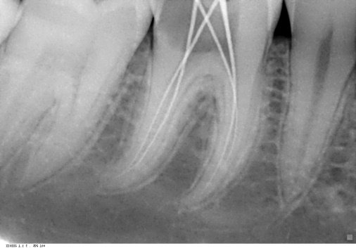
PRE-OPERATIVE & WORKING LENGTH
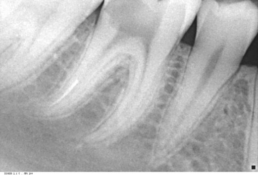
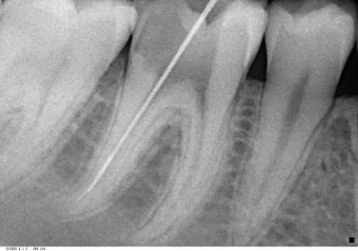
FILE SEPARATED & BYPASS DONE

MASTER CONE (MESIAL VIEW), MASTER CONE (STRAIGHT VIEW), & MASTER CONE (DISTAL VIEW)
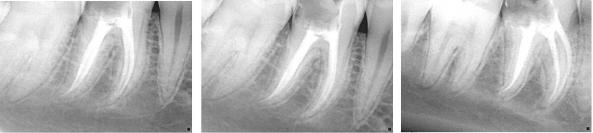
OBTURATION (MESIAL VIEW), OBTURATION (STRAIGHT VIEW), OBTURATION (DISTAL VIEW)
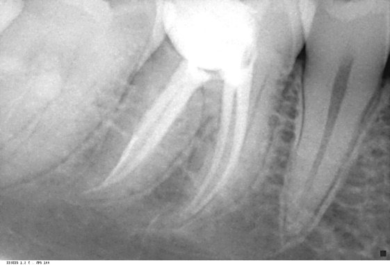
POST ENDODONTIC RESTORATION
CASE 02:
A 21 year old male patient reported with a chief complaint of continuous and radiating pain in relation to lower left back tooth region since a week. The patient also complained of episodes of sensitivity to hot and cold in the involved tooth. Clinical examination revealed left mandibular first molar with proximal caries on the distal aspect. Upon Radiographic examination, radioleucency in the crown involving pulp and widening of the pdl space was observed. Based upon the clinical and radiographic examination, diagnosis of Chronic Irreversible Pulpitis with Apical Periodontitis w.r.t 36 was made and treatment plan was Non Surgical Root Canal Treatment w.r.t 36. After administration of local anesthesia, tooth was isolated, pre-endodontic buildup was done, a straight line endodontic access cavity was made. During cleaning and shaping, F1 Rotary file was separated in mesiobuccal canal of the tooth. A radiograph was taken to confirm the level of separation of the instrument. The instrument was found to be separated at the apical 3rd of the distal canal. A nonsurgical file bypass technique was selected for this case. #8K file was inserted between the fracture fragment and the root canal wall, negotiating the canal to full working length which enables thorough instrumentation. The patency of the canal was found with #10K, #15K, #20K, #25K in that position, a working length measuring radiograph was recorded. Calcium hydroxide dressing was given for 7 day. Obturation was done and Final radiograph was taken.
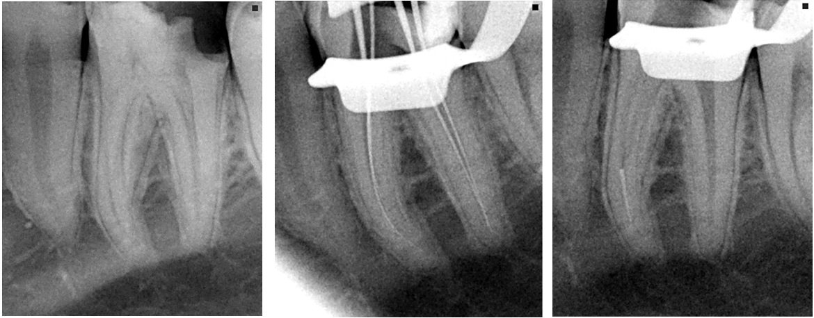
PRE-OPERATIVE, WORKING LENGTH, & FILE SEPARATED

BYPASS DONE, MASTER CONE, & OBTURATION
DISCUSSION
Separated instruments can be handled conservatively or surgically. The conservative methods such as leaving the fragment in situ and filling the RC to the coronal level of the instrument, bypassing the segment or removing it from the RC are considered favorable and preferred whenever possible.1
When an instrument fracture occurs, the clinician needs to evaluate the separated portion radiographically and clinically.3 Then find the treatment options with consideration for the pulp status, root canal infection, root canal anatomy, position, size and type of fractured instrument and the amount of damage that would be caused to the remaining tooth structure.4
A technique that does not require direct visibility to the fragment is “bypass”, where a fine file is inserted between the fragment and root canal wall and thereby negotiating the canal to full working length and enable thorough instrumentation and obturation with the fragment remaining in situ. Incorporating the fragment in the root canal obturation material considerably improves the case prognosis.5
BYPASSING
“Bypassing technique” based on the fact that none of the root canals are perfectly round, and a small gap exists between the root canal wall and the fractured fragment, which allows a smaller file to bypass the separated fragment. It involves inserting a fine file between the fracture fragment and the root canal wall, and negotiating the canal to full working length which enables thorough instrumentation and root canal obturation with the fragment remaining in situ. Allowing the fragment in situ along with thermo plasticized guttapercha considerably improves the prognosis. This is an easy technique to master, and works out successfully majority of times, especially when the instrument is bound in coronal and middle thirds of canal.
While bypassing with the fine or small size instrument, there can be a possibility that the fractured segment can be retrieved from the canal. One more advantage of this technique is it does not demand direct visibility to the fragment i.e. it can be suitable when the fragment is located beyond a considerable root canal curvature. This method does not demand magnification aids strictly, as it is more dependent on tactile sensation of dentist, allowing its practical feasibility among general dentists.2
INDICATIONS
For fragments located in the middle or apical 1/3 of the RC;
- For fragments beyond the curvature
- After failure of a previous removal attempt
- In cases when perfect visualization and straightening of the RC cannot be achieved or would be too structurally invasive.
ADVANTAGES
- More conservative procedure than the complete removal of the fragment
- Less invasive
- Relatively simple procedure
- Often results in complete removal of the fragment
- Allows achieving full working length, proper preparation, irrigation and obturation of the RCS.
RISKS
- Possibility to create a false RC, parallel to the original one
- Ledge formation
- Possible perforations
- Possible secondary separation of instruments
- Possible fragment extrusion trough the apical foramen1
MEDICO-LEGAL CONSIDERATIONS
It is considered good practice for the clinician to inform the patient preoperatively of the options and risks of treatment as part of the consent procedure. Theoretically therefore, every patient undergoing endodontic treatment or retreatment should be warned of the possibility of file fracture. However, it is questionable as to whether this happens in practice.
The incidence of instrument fracture is reported to be relatively low (0.7-7.4%) and the effect of instrument fracture on outcome is largely reported as insignificant. Nevertheless, the stressful event of fracture becomes more difficult to defend and manage if the patient has not been informed of the potential risk or indeed of the presence of a fractured instrument in their root canal. What is certain is the fact that clinicians have a legal obligation to inform the patient - and document it in the patient’s notes - if an instrument has fractured during treatment.6
CONCLUSION
Instrument separation can happen even if the instrumentation protocol was followed precisely. The decision for conservative management to bypass or to completely remove the fractured instrument should be made after careful evaluation of various factors, such as root canal system anatomy and mor- phology, exact location of the fragment, presence or absence of periapical pathology and armamentarium available. From the two cases mentioned in this article we were able to reach to a conclusion that if the separated instrument is able to be bypassed, it is more conservative than retrieving the separated instrument.
REFERENCES
- Tsvetelina Borisova-Papancheva, Silvia Stankova, Slavena Georgieva. Conservative Management Of Intracanal Separated Endodontic Instruments: Treatment Decisions And Related Factors. Scripta Scientifica Medicinae Dentalis, Vol. 3, No 1, 2017, Pp. 23-31.
- Rambabu T. Management Of Fractured Endodontic Instruments In Root Canal: A Review. Journal Of Scientific Dentistry 2014;4(2):40-48
- Dr Rafaël Michiels,. Bypassing A Fractured Instrument. Case Report. Roots1_2011.
- Sultana Parveen, Mozammal Hossain And Md. Farid Uddin. Management Of Broken Instrument By File Bypass Technique. Bsmmu J 2017; 10: 41-43
- Dr. Praveen John, Dr. Ramesh Kumar M., Dr. Jayasree S. Bypassing Separated Instruments In The Root Canal – Two Case Reports. IOSR Journal Of Dental And Medical Sciences (Iosr-Jdms) Volume 14, Issue 7 Ver. Xi (June. 2016), Pp 08-13.
- M. B. Mcguigan, C. Louca And H. F. Duncan. Clinical Decision-Making After Endodontic Instrument Fracture. British Dental Journal Volume 214 No. 8 Apr 27 2013.
Disclaimer from Guident
The views and opinions expressed in articles published in Guident are solely those of the authors and do not necessarily reflect the views of the editors, the editorial board or the publisher, Ivory India. Guident does not endorse any product, technique or treatment described. Clinical content is intended for qualified dental and medical professionals and is not a substitute for individual clinical judgement. Guident, its editors and the publisher accept no liability for any consequence arising from the use of information published in the journal.
How to cite this article
Dr. Sneha, Dr. Annil Dhingra. Conservative Management of Intracanal Separated Endodontic Instruments Using File Bypass Technique- a Case Series. Guident. 2020 Dec;14(1). Available from: https://guident.in/articles/endodontics/conservative-management-of-intracanal-separated-endodontic-instruments-using-file-bypass-technique-a-case.html


