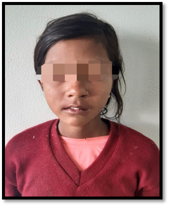
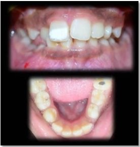
Evidence Based Treatment Planning for Dentigerous Cysts
Department of Pediatric and Preventive Dentistry, Manipal College of Dental Sciences, Mangalore, Manipal Academy of Higher Education, Manipal
ABSTRACT
Introduction: Dentigerous cysts (DCs) are one of the most commonly seen developmental odontogenic cysts. They occur more frequently in the second and third decade of life. DCs enclose the crown of the impacted tooth, at the cervical region and originate from the epithelial remnants of the tooth-forming organs. A strong association is known to exist between the failure of eruption of the tooth and the dentigerous cyst. Case reports: Hereby, we present 2 cases of dentigerous cysts and their surgical management. Case 1: A 10-year-old boy reported with a chief complaint of swelling in the lower left back region. Clinical examination showed that the patient was in the late mixed dentition phase, with a buccal hard tissue swelling in the region of 34, 35. The radiographic and CBCT examination revealed a well circumscribed unilocular radiolucency, causing a breach of the cortical plate in the third quadrant (34, 35). The treatment consisted of enucleation of the cyst along with the removal of the affected tooth. Case 2: A 7 year-old young boy reported with a swelling in the upper right front region. Clinical examination revealed an early mixed dentition phase, with a complete obliteration of the buccal vestibule in relation to 53. Radiographic evidence showed the presence of an ill-defined radiolucency in the canine region. The CBCT revealed a well-defined expansile lesion suggestive of a dentigerous cyst. The treatment comprised of enucleation of the cyst and removal of 53. Recall: Both patients are asymptomatic and have shown no reoccurrences.
INTRODUCTION
A cyst is defined as a pathologic cavity containing fluid, semifluid or gaseous contents that is not created by the accumulation of pus; and is frequently but not always, lined by epithelium. Odontogenic cysts (OC) can be arbitrarily classified into two groups based on their origin: Developmental and inflammatory. Odontogenic cysts cause the resorption of bone and develop from the components of the odontogenic epithelium or its residuals.1 Dentigerous cysts (DCs) are the second most commonly seen developmental odontogenic cysts, with a male to female ratio of 1.17:1 in India. They occur more frequently in the second and third decade of life. Mandibular posterior regions are the most frequently affected sites (53.85 %) followed closely by the maxillary posterior region (23.08 %).2 DCs enclose the crown of the impacted tooth, at the cervical region and originate from the epithelial remnants of the tooth-forming organs. A strong association is known to exist between the failure of eruption of the tooth and the dentigerous cyst.3
The current treatment protocol for Dentigerous cysts comprises of surgical enucleation and extraction of the affected tooth.1 However, enucleation of the cyst, while retaining the affected permanent tooth is also a viable option that can be considered based on the age of the child. Fujii et al. and Yahara et al, stated that the spontaneous eruption of the affected tooth is possible in patients < 10 years old, provided that the depth of inclusion is less than 5.1 mm and that tooth angulation is smaller than 25°.4 This case series presents two such cases, where in two different treatment strategies were used.
CASE 1
A boy aged 10 years reported to the dental out patient with the chief complaint of a swelling in the lower left back tooth region since the past one week. The swelling had presented itself before and the patient had gone to another clinic, where they had been prescribed anti-inflammatory analgesics. The swelling had subsided but reappeared within a week. The family gave no other relevant medical history. Upon visual examination of the child, an extra-oral facial swelling resulting in facial asymmetry was seen. The swelling extended from the angle of the mandible to the nasio-labial fold antero-posteriorly and from the lower border of the mandible to the infra-orbital margin supero-inferiorly (FIGURE 1). Palpation revealed a bony hard swelling intra-orally (2.5cms in M-D width and 1cm superior-inferior width) in the region of the primary first and second left molar. There was an over-retention of the first primary molar, with the presence of an erupting first premolar (FIGURE 2). The provisional diagnosis arrived at was that of a Dentigerous Cyst.
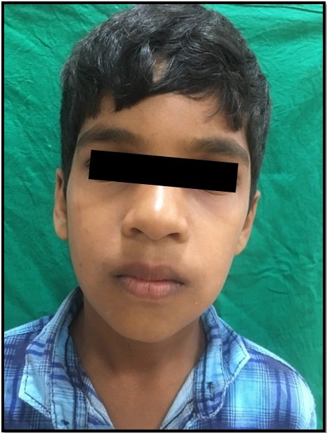
Figure 1: Extra-Oral Swelling on the lower left region

Figure 2: Over retained 74, seen with erupting 34. Presence of a bony hard swelling measuring 2.5X1cm
The OPG and CBCT confirmed the presence of a well circumscribed radiolucency with respect to the second premolar, showing a breach of the cortical plate and proximity to the nerve canal. (FIGURE 3) Based on the article by Hyomoto et al. the cusp depth of the impacted tooth from the alveolar crest and the angle formed between the long axis of the impacted tooth and the long axis of adjacent teeth, were assessed. The depth of inclusion and angulation was greater than 5mm and 50° respectively.4
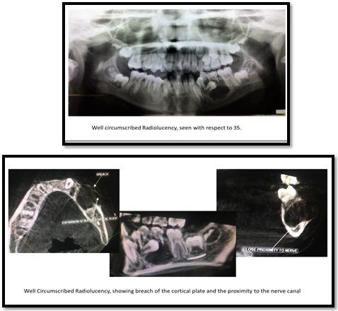

Figure 3: OPG, Focal CBCT, Parameters Measured
The treatment plan formulated for the case consisted of surgical enucleation of the cyst, with extraction of the primary first molar and second premolar. (FIGURE 4) The excisional biopsy report revealed a soft tissue lesion that was white to brown in colour, smooth to rough and soft to firm in consistency. It measured 2 x 1 x 0.5cms in size. The microscopic appearance showed the presence of an inflamed connective tissue capsule lined intermittently by 2-5 cell layers of flattened, non-keratinized stratified squamous epithelium. The connective tissue capsule was moderately dense, showing inflammatory cells. Hence the report confirmed the diagnosis of an Inflamed Dentigerous Cyst. The child was called back for a follow up 10days (FIGURE 5) and 1month post the operation. He was asymptomatic and the healing was uneventful.

Figure 4: Surgical Enucleation of the Cyst

Figure 5: 10 days Post-Operative Pictures
CASE 2
A 7 year old boy reported to the clinic with a chief complaint of intra-oral swelling in the upper right back tooth region since past 1 month. There was no history of any pain or associated sensitivity. Upon examination, an intra-oral swelling measuring 2*2 cm in the sulcular region in relation to the upper right primary canine was noticed. There was also an obliteration of the buccal vestibule and the buccal cortical plate was soft on palpation. (FIGURE 6) The provisional diagnosis arrived at was that of a Dentigerous Cyst.

Figure 6: Pre-operative Intra-Oral Pictures
The OPG and CBCT revealed the presence of a well-defined expansile radiolucent lesion (18*22mm) in the canine region with the deflection of the permanent lateral incisor and canine tooth buds. (FIGURE 7)

Figure 7: OPG and CBCT revealing well defined radiolucency in the region of 53
In this case, the dentigerous cyst was enucleated and the affected permanent tooth was not extracted. (FIGURE 8) The child was asymptomatic, and the healing was uneventful at the 1 month follow up. At the 2 year follow up, the permanent first premolar had erupted albeit ectopically, but the child was asymptomatic

Figure 8: Enucleation of cyst, extraction of primary canine
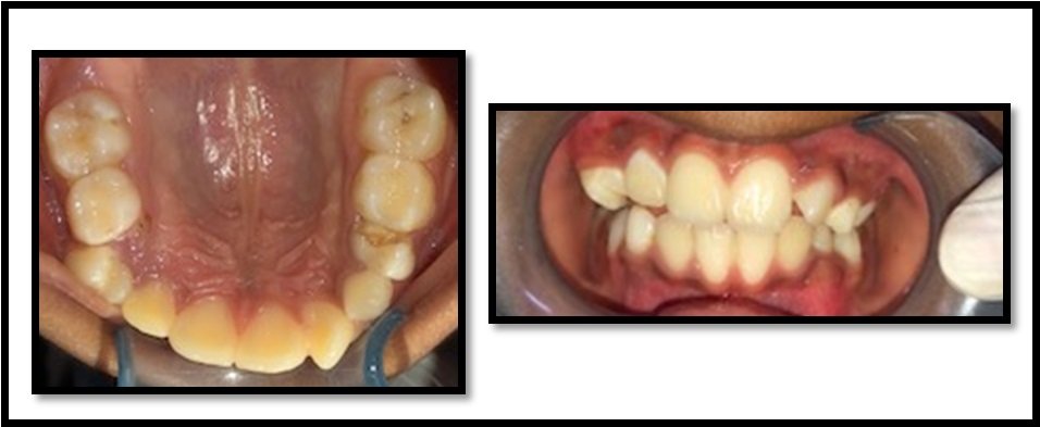
Figure 9: 2 year Follow up Pictures
DISCUSSION
Dentigerous cysts are Odontogenic Developmental Cysts, that show a male predilection. They occur due to the accumulation of fluid between the reduced enamel epithelium and the crown of the tooth.3 The exact etiological cause for the occurrence of the cyst is unclear, but an infection of the primary tooth can be a plausible factor for the formation of a dentigerous cyst with respect to the underlying permanent tooth bud. The cyst is characterized by a slow growing bony hard swelling of the jaw, with the absence of the affected tooth in the dental arch. The patient usually displays facial asymmetry, with cortical plate expansion and perforation (in some cases).3 Histopathologically, dentigerous cysts reveal stratified squamous non-keratinized epithelium, that are usually 2-3 cell layers thick. In cases, that the dentigerous cysts are left undiagnosed, the cysts may transform into Ameloblastomas. Hence the early detection and treatment of dentigerous cysts is imperative.3
The treatment modalities that exist for Dentigerous cysts include enucleation or marsupialization. Enucleation is defined as the surgical removal of the cyst, without cutting or dissecting it, along with the affected tooth.4 Marsupialization on the other hand, involves allowing the cystic fluid to drain, and the lining to shrink, before curetting it out from the site. Both procedures have their own pros and cons.5 The rate of recurrences and discomfort to the patient are reduced, in enucleation, whereas marsupialization displays bone reparative capacity, with the potential of immature teeth to spontaneously erupt after the removal of the cyst.4,5 In the present cases, both children were treated with enucleation so as to eliminate the need for open drainage.5
Hyomoto et al. evaluated multiple parameters to assess the chances of spontaneous eruption of the impacted tooth. They measured the cuspal depth of the impacted tooth, the angle formed between the long axis of the impacted tooth and the long axis of adjacent teeth, root maturity, cystic area, and the eruption space available.5 They concluded by saying eruption was witnessed in cases where in the depth of inclusion was less than or equal to 4.4 mm and the angulation of the tooth was 20.4° ± 21.8°.5 In similar articles by Fujii et al. in 2008 and Yahara et al. a parallel was drawn between the age of the patients and the chances of spontaneous eruption. The articles stated that if the child was below 10years of age (mean age 9.8years), with the eruption space present larger than 1 cm, and the root incompletely formed, the tooth had a higher chance of spontaneous eruption.5
Hence, in the first case since the child had completed 10years of age, with the depth of inclusion and angulation greater than 5mm and 50° respectively, the chances for the tooth to re-erupt was poor, and the decision to extract the tooth was taken. In the second scenario, the child reported to the clinic at 7 years of age, with sufficient space for the canine to erupt and an incompletely formed root apex. Therefore, the permanent canine was retained.
CONCLUSION
Dentigerous cysts, are a common prevalence, that require proper clinical and radiographical analysis. This not only helps clinicians arrive at an accurate diagnosis but also assists in the formulation of a suitable treatment modality.
REFERENCES
- Rajendra Santosh AB. Odontogenic Cysts. Dent Clin North Am. 2020;64(1):105-119.
- Kambalimath DH, Kambalimath HV, Agrawal SM, et al. Prevalence and distribution of odontogenic cyst in Indian population: a 10 year retrospective study. J Maxillofac Oral Surg. 2014;13(1):10-15.
- Purkait, S.K. Essentials of Oral Pathology. Third edition. New Delhi, India: Jaypee Brothers Medical Publishers. 2011.
- Shetty RM, Dixit U. Dentigerous Cyst of Inflammatory Origin. Int J Clin Pediatr Dent. 2010;3(3):195-198.
- Ghandour L, Bahmad HF, Bou-Assi S. Conservative Treatment of Dentigerous Cyst by Marsupialization in a Young Female Patient: A Case Report and Review of the Literature. Case Rep Dent. 2018; 2018:7621363.
Disclaimer from Guident
The views and opinions expressed in articles published in Guident are solely those of the authors and do not necessarily reflect the views of the editors, the editorial board or the publisher, Ivory India. Guident does not endorse any product, technique or treatment described. Clinical content is intended for qualified dental and medical professionals and is not a substitute for individual clinical judgement. Guident, its editors and the publisher accept no liability for any consequence arising from the use of information published in the journal.
How to cite this article
Dr. Deveshi Gupta, Dr. Sowmya Sridhar, Dr. Arathi Rao. Evidence Based Treatment Planning for Dentigerous Cysts. Guident. 2020 Dec;14(1). Available from: https://guident.in/articles/pedodontics/evidence-based-treatment-planning-for-dentigerous-cysts.html