Confirmation of Radiographic Diagnosis of Supernumerary Roots Using Cone Beam Computed Tomography
Department of Conservative Dentistry & Endodontics, Post Graduate Institute of Dental Sciences, Rohtak.
ABSTRACT
Anatomical variations like the supernumerary roots are an acknowledged characteristic in permanent molars and can pose great challenges to the clinician. Radiographs aid in identiï¬cation and assessment of supernumerary roots. But they are two dimensional view of a three dimensional tooth structure. Cone Beam Computed Tomography (CBCT) images provide a better understanding of root canal anatomy aiding in an accurate detection of supernumerary roots thereby avoiding complications in endodontic treatment. This case report highlights the importance of CBCT in confirming an uncertain radiographic diagnosis of supernumerary roots.
INTRODUCTION
The main objective of root canal treatment is the thorough mechanical and chemical cleaning of the entire pulp space followed by complete obturation with inert filling material. Therefore, it is imperative that aberrant anatomy is identified before and during root canal treatment. Radiographic examination forms an essential and the most common preoperative assessment tool for root canal treatment. However, the conventional two dimensional radiographs might not yield sufficient diagnostic information. Three dimensional imaging overcomes the major limitations posed by the radiographs by allowing the visualization of the root morphology in all the three parameters i.e. transverse, axial, and sagittal.
Mandibular first molars are the most commonly encountered teeth requiring root canal treatment. These teeth display several anatomical variations which are thought to be racially and genetically determined.1 A high incidence of third root, Radix Entomolaris (RE) (distolingually) / Radix Paramolaris (RP) (mesiobuccally), is identified as a typical variation in lower first molars.2 Therefore, a clinician must always be aware of the presence of an extra root while treating a mandibular first molar.
Clinical analysis of the tooth crown and the cervical morphology of the roots by means of periodontal probing can help identify an additional root.1 An extra cusp (tuberculum paramolare) or more prominent occlusal distal or distolingual lobe with a cervical prominence, indicate the probability of an extra root.1
Radiographic examination has been regarded as a suitable procedure for preoperative identification of RE. But the conventional radiography has its own shortcomings. Unseen curvatures on orthoradial radiographs may account for loss of working length, canal transportation and strip perforation .3 Also the double outlining seen on conventional radiography may not always correctly point out the presence of an extra root and may lead to a wrong diagnosis of RE. In view of these shortcomings of the conventional radiography, the non-invasive three-dimensional imaging technique, Cone Beam Computed Tomography (CBCT), provides another choice for further assessment. Using CBCT, seven different categories of variants in the root canal anatomy of mandibular first molars have been recognised by Zhang et al., out of which variant 5 and 6 correspond to three separate roots and variant 7 to four separate roots.4
The present case report highlights how the conventional radiography may lead one to a paradoxical diagnosis of anatomical aberrations by giving a two dimensional view of a three dimension structure; and the importance of three dimensional imaging using CBCT for a confirmatory diagnosis of supernumerary roots.
CASE REPORT 1
A 45 yrs old male patient reported with a chief complaint of recurrent painful episodes in lower left molar region. Clinical examination showed an occlusal restoration in 36. The tooth was sensitive to percussion, thermal and electric pulp tests. Radiograph at 0 degree angulation showed signs of apical periodontitis, loss of lamina dura with radiolucent changes in the furcation area of 36 (Fig. 1). The radiograph revealed a double periodontal ligament lining of both the mesial and the distal roots. To confirm the findings another radiograph was taken at a 25 degree mesial angulation (Fig. 2). The periodontal ligament linings in the second radiograph were distinctly seen to be distantly apart in both the roots.
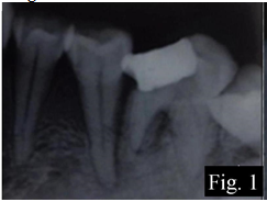
Fig. 1: IOPA radiograph of case 1 showing double outlining of both mesial and distal roots at 0 degree horizontal angulation.
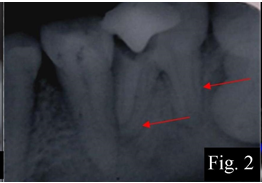
Fig. 2: IOPA radiograph of case 1 at a 25 degree mesial angulation.
Radix generally presents with a crossing over of the periodontal ligament linings which was lacking in both the preoperative radiographs. Thus, a CBCT scan to have a definitive diagnosis of the root and canal anatomy was done. The transverse section of the tooth showed two separate roots only with two canals in the mesial and two canals in the distal root (Fig. 3). This refers to Variant 4 as described by Zhang et al.
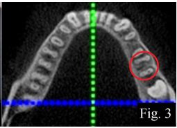
Fig. 3: CBCT of case 1
After anaesthesia and rubber dam isolation an endodontic access cavity was prepared and all the four canal orifices were identified. The root lengths were estimated using an apex locator (Root ZX; Morita, Tokyo, Japan) and then confirmed with a periapical radiograph. All canals were instrumented following step back technique with intermittent irrigation using 5.25% sodium hypochlorite. A final irrigation was performed alternating 17% ethylenediaminetetraacetic acid and 4.2% NaOCl solution. The root canals were then flushed with sterile saline and dried using sterile paper points. After preparation, the canals were obturated with lateral compaction technique. A permanent restoration was given following endodontic therapy.
CASE REPORT 2
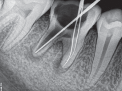
A 21 years old male reported with complaints of pain on chewing in left lower back region. Clinically, a grossly decayed mandibular left first permanent molar (36) was seen. The tooth was sensitive to percussion, thermal and electric pulp testing. A preoperative radiograph at 0 degree angulation revealed an exposed distal pulp horn and a widened periodontal ligament space around the roots with loss of lamina dura (Fig. 4). The radiograph revealed a double root outlining in both the mesial and distal roots. This raised a suspicion of additional roots in the molar. Subsequently, another radiograph was taken at 25 degree mesial angulation (Fig. 5). A double periodontal ligament lining around both the mesial and the distal roots was seen farther apart as compared to the previous radiograph. But a distinct impression of extra roots with extra canals was not visible on both the radiographs taken at different angulations. After due consent of the patient, further confirmation was done using CBCT. The transverse cross-section of CBCT revealed Zhang’s variant 4 type root anatomy (Fig. 6). Further treatment plan was as described in case 1.

Fig. 4: IOPA radiograph of case 2 at 0 degree angulation showing double outlining of both mesial and distal roots.
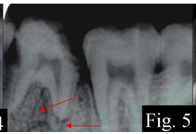
Fig.5: IOPA radiograph of case 2 at 25 degree mesial angulation further giving an impression of 4 roots with 4 canals
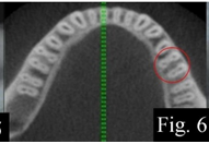
Fig. 6: CBCT of case 2
CASE REPORT 3
A 15 years old male reported with a chief complaint of pain in right lower back tooth since one month. Clinical examination revealed a grossly carious mandibular right first molar. The tooth was sensitive to percussion and thermal testing. Preoperative radiographs (0 degree and 25 degree) showed widened periodontal ligament space (Fig. 7). A diagnosis of irreversible pulpitis was made. Radiographs also revealed a clearly identifiable distolingual root image (Wang’s type i). An additional diagnostic evaluation in terms of CBCT could not be advocated. Further treatment plan was as in case 1.
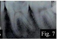
Fig. 7: IOPA radiograph of case 3 at 25 degree horizontal angulation distinctly showing a distolingual root.
DISCUSSION
The presence of RE / RP has clinical implications in endodontic treatment and a special attention to the extra roots in mandibular molars is necessary.
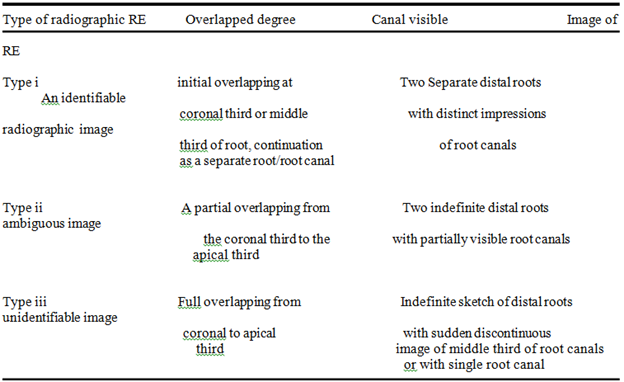
On the basis of the radiographic overlapped degree between distolingual and distobuccal root, the radiographic features of RE were classified into 3 types by Wang et al (Table 1): type i, slight overlapped image; type ii, moderate overlapped image of RE; type iii, severe overlapped image of RE (3). Since RE is generally situated in the same bucco-lingual plane as the distobuccal root, a pre-operative radiograph may not be able to rule out its presence every time. An angled view may be helpful in some cases. Studies have found that a 25-degree mesial radiograph is significantly better than a 25-degree distal radiograph for an optimum diagnosis of anatomical structures.3,5
In our case reports, radiographic image of case 1 simulated Wang’s type ii i.e. moderately overlapped at 0 degree exposure. Further angulating the beam at 25 degree revealed distant periodontal ligament linings along the roots creating an impression of supernumerary roots. Similarly in case 2, Wang’s type iii i.e. severely overlapped images of tooth root were seen. At 25 degree angulation a distinct periodontal lining along both the roots was separately seen. Yet in both the cases an identifiable radiographic proof giving a distinct impression of separate roots with separate canals was lacking. Thus, in both the cases a CBCT scan was planned for confirmatory diagnosis of supernumerary roots. Although radiographs created an ambiguous situation, absence of any supernumerary roots was confirmed from the scan images. The double root outlining initially seen on the radiographs could be attributed to the dumble shaped root anatomy.
TABLE 1 Radiographic Features of Radix Entomolaris (RE)
Whereas in case 3, the radiographic image of an extra distolingual root was clearly appreciable at 25 degree mesial angulation (type i), therefore, CBCT evaluation was not done as it would mean an unnecessary radiation exposure to the patient.
Identification of all relevant clinical anatomies is not guaranteed even by multiple intraoral radiographs. The combination of sagittal, coronal and axial CBCT images gives the clinician a more in-depth understanding of the true morphology of root canal systems. Recently, a few authors have corroborated the use of CBCT to be included as a standard diagnostic means for a successful outcome of endodontic therapy. 6,7 Zhang et al have divided the root canal anatomy of mandibular first molars into seven different variants using CBCT (Table 2).
CBCT has limitations related to the ‘‘cone-beam’’ projection geometry, detector sensitivity, contrast resolution and scattered radiations.8 But still CBCT is a good tool for studying intricate details
TABLE 2

of root canal morphology for endodontic treatment provided each radiation exposure is justified. Therefore, it is sensible to adopt CBCT for further diagnostic evaluation only when there are abnormal and inconclusive findings on traditional periapical films.
REFERENCES
1. Abella F, Patel S, Durán-Sindreu F, Mercadé M. Mandibular first molars with disto-lingual roots: review and clinical management. Int Endod J. 2012; 45(11): 963-78.
2. Calberson FL, De Moor RJ, Deroose CA. The radix entomolaris and paramolaris: clinical approach in endodontics. J Endod. 2007; 33(1): 58-63.
3. Wang Q, Yu G, Zhou XD, Peters OA. Evaluation of x-ray projection angulation for successful radix entomolaris diagnosis in mandibular first molars invitro. J Endod. 2011; 37(8): 1063-8.
4. Zhang et al. Use of cone-beam computed tomography to evaluate root and canal morphology of mandibular molars in Chinese individuals. Int Endod J. 2011; 44(11): 990–999.
5. Fava LR, Dummer PM. Periapical radiographic techniques during endodontic diagnosis and treatment. Int Endod J. 1997; 30(4): 250–61.
6. Estrela C, Bueno MR, Leles CR, Azevedo B, Azevedo B, Azevedo JR. Accuracy of cone beam computed tomography and panoramic and periapical radiography for detection of apical periodontitis. J Endod. 2008a; 34(3): 273–9.
7. Wu M-K, Shemesh H, Wesselink PR. Limitations of previously published systematic reviews evaluating the outcome of endodontic treatment. Int Endod J. 2009; 42(8): 656–66.
8. Sogur E, Baksi BG, Grondahl HG. Imaging of root canal fillings: a comparison of subjective image quality between limited cone beam CT, storage phosphor and film radiography. Int Endod J. 2007; 40 (3): 179–85.
Disclaimer from Guident
The views and opinions expressed in articles published in Guident are solely those of the authors and do not necessarily reflect the views of the editors, the editorial board or the publisher, Ivory India. Guident does not endorse any product, technique or treatment described. Clinical content is intended for qualified dental and medical professionals and is not a substitute for individual clinical judgement. Guident, its editors and the publisher accept no liability for any consequence arising from the use of information published in the journal.
How to cite this article
Dr. Meenu Dhiman Dr. Jigyasa Duhan Dr. Alpa Gupta. Confirmation of Radiographic Diagnosis of Supernumerary Roots Using Cone Beam Computed Tomography. Guident. 2014 Jun;7(7). Available from: https://guident.in/articles/endodontics/confirmation-of-radiographic-diagnosis-of-supernumerary-roots-using-cone-beam-computed-tomography.html