Prosthodontic Rehabilitation of a Patient with Kennedys Class 1 Using Fixed Partial Denture and Cast Partial Denture with Precision Attachments: A Case Report
Department of Prosthodontics and Crown & Bridge, Sardar Patel Post Graduate Institute of Dental and Medical Sciences, Lucknow, Uttar Pradesh, India
ABSTRACT
A prosthesis delivered should cater to the needs of the patient and also meet the biological, mechanical, and esthetic considerations. Care should be taken in the formulation of treatment plan that the planned prosthesis fulfills the requirements of the patient and the dentist. Among the various methods of oral rehabilitation precision attachments are viable alternative to join fixed (FDP) and cast partial dentures (CPD) and thereby avoiding cumbersome procedures. The advantages of attachment retained CPD are the improvements in esthetics which eliminates the clasp assembly and biomechanics. This article describes the treatment sequence and technique for the use of attachments in therapy of combining FDP/CPD of patient reported with dislodged cantilever fixed prosthesis.
Keywords:- precision attachment, CPD, FPD, OT cap , rhein 83
INTRODUCTION
Esthetically and functionally successful prosthetic rehabilitation requires careful attention and meticulous treatment planning1. Rehabilitation of partially edentulous arch can be challenging when it is a distal extention situation classified under Kennedy’s class I and class II situations. A fixed partial denture cannot be created in this situation due to the absence of the distal abutment. Although implant-supported prostheses can be planned, there are situations when they are not practical for medical or financial reasons. So, in such situation an acrylic partial denture or a cast partial denture is largely preferred. Cast partial dentures are made retentive by the use of retainers and precision attachment components. Precision attachments could be extracoronal and intracorona.l Attachment-retained cast partial dentures facilitate both esthetic and functional replacement of missing teeth.2 The available restorative space along with the number of teeth missing and the periodontal health of the abutment teeth pose a significant challenge while determining the design and fabrication of specific RPD attachments for extracoronal fittings, a modified extended precision attachment system to achieve optimum retention, stability, and enhancing the esthetics by eliminating the visible metal components.3 Precision attachment is an interlocking device, one component of which is fixed to an abutment or abutments, and the other is integrated into a removable dental prosthesis in order to stabilize or retain it. These attachments take the place of damaging clasp arm. Based primarily on the function of these attachments they are classified as rigid or passive. OT CAP Attachment (rhein 83inc, USA) a passive type of attachment consists of a matrix (a sphere available in preformed plastic patterns, which is cast to the abutments and a patrix which are preformed housings which are casted into the framework and contains retentive caps made of nylon or rubber in different colures corresponding to different retentive degrees, both in normal and micro size.4
CASE DESCRIPTION
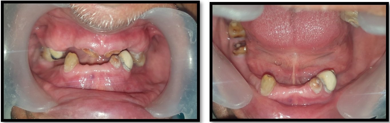
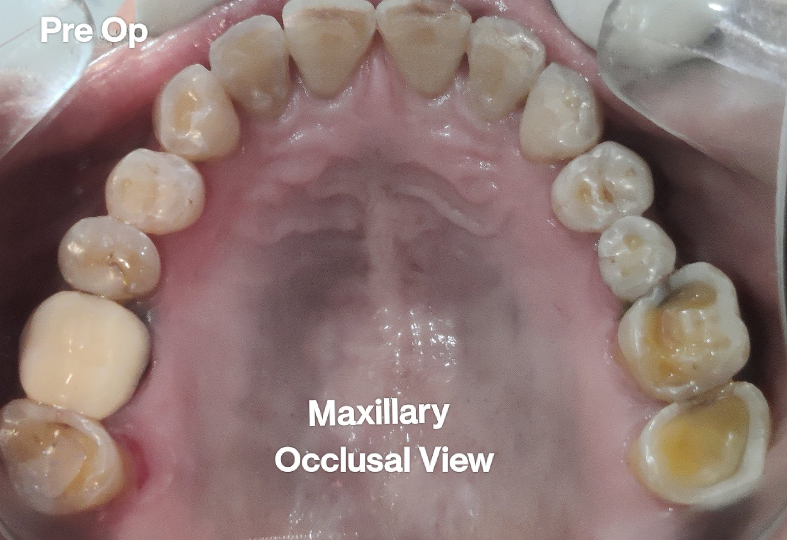
A 58-year-old male patient, referred from the Department of Oral Medicine and Radiology to the Department of Prosthodontics and Crown and Bridge, Sardar Patel Post Graduate Institute of Dental and Medical Science, Lucknow, Uttar Pradesh, India, for the replacement of missing teeth. Patient’s chief complaint was that he had lost the teeth due to caries and had been partially edentulous for the past 2 years He complains of an unappealing, unattractive appearance and difficulty in chewing food due to the missing teeth in his upper and lower posterior region. On clinical examination, teeth present were 11, 12, 13, 15 and 21 in the maxillary arch and 42, 43, 44, 32, 33, 34, 35 and 36 in the mandibular arch. , and a dislodged porcelain fused to metal crown with 35, 36, 43, 44 was present.

Fig 1: pre-operative Intraoral Picture.
Clinical and radiographic examination revealed the presence of generalized attrition and reduced vertical dimension of occlusion, which was reduced due to the loss of maxillary and mandibular posterior teeth .The patient was diagnosed as a favorable partially edentulous case for full-mouth rehabilitation as Kennedy’s class I in the maxillary and Kennedy’s class I mod I in mandibular arch, and 32 and 36 were extracted due to grade 3 mobility. The treatment option suggested was the precision attachment retained cast partial denture in the maxillary and mandibular arch with intentional endodontic therapy was performed in all the remaining teeth.
LAB PROCEDURES
After complete clinical and radiographic examination, a prosthetic treatment plan was set up. Combined prosthesis with extracoronal precision attachment was planned for maxillary and mandibular bilateral distal extension. Tooth preparation of abutment teeth was performed to receive porcelain fused to metal crowns. The abutments prepared were temporized after making definitive impression.5
- Waxing up of abutments was performed and milling of lingual area of metal ceramic setup was done. Articulation spaces and bulkiness were evaluated in order to proceed with optimal positioning of attachments using proper parallelometer mandrel.
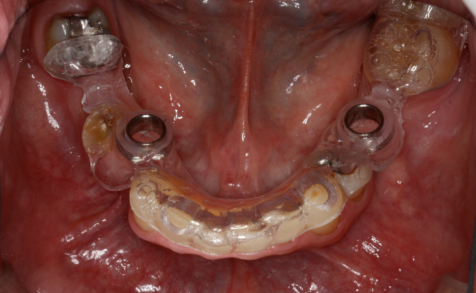
- Metal ceramic crowns waxed up with attachment structure were casted and porcelain firing was done. Splinted crowns were fabricated with the attachments in the laboratory and the trial of the same was done to check the exact fit of the crowns.

Fig 2: Framework fabricated of FPD with precision attachment.
- Cast partial denture with attachment was fabricated in the laboratory and the metal framework trial was done in the patient’s mouth for the accuracy of fit, Cast structure framework was checked up for stability and precision and jaw relation were recorded.6

Fig 3: Try-in of metal framework on both arches.
- Waxing up of teeth was performed and teeth setting trial was done in patient’s mouth. The trial denture was sent for acrylization and cast partial denture finished.

Fig 4: Try-in done with CPD and FPD (teeth arrangement on wax ) and occlusion checked.
- Trial seating of the finished prosthesis was performed and cementation of crowns was done using Glass Ionomer cement (GC Fuji). Attachments are protected with a thin layer of petroleum jelly (Vaseline) in order to easily remove cast partial denture after joint PFM crowns with attachment have been seated. Complete seating of finished maxillary and mandibular combined prosthesis with extracoronal castable distal extension precision attachment was evaluated clinically and the patient was recalled after 24 hrs for post-insertion checkup.7

Fig 5: Final prosthesis with precision attachment (maxillary).

Fig 6: Final prosthesis with precision attachment (mandibular).
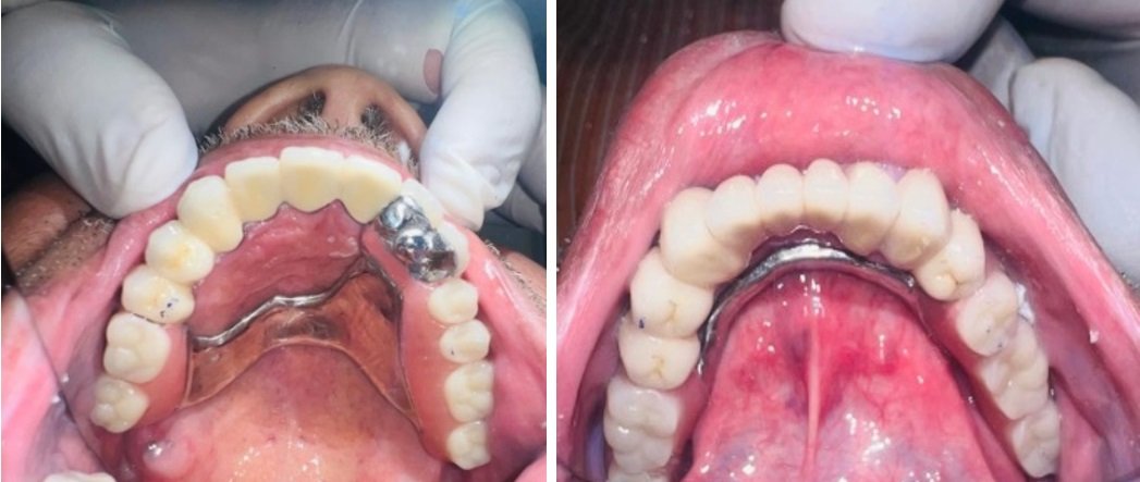
Fig 7: Delivered precision attachment prosthesis with FPD.


Fig 8: post-op intraoral pictures.
DISCUSSION
Precision attachment is a connector consisting of two or more parts. One part is connected to a root, tooth, or implant and the other part to the prosthesis providing a mechanical connection between the two. These attachments allowed prosthesis to combine the advantage of fixed and removable restorations.8,9,10 It was Dr. Herman Chayes who first reported the invention of attachment in the early 20th century. The stress on the abutment due to the difference in nature and behavior of the tissues supporting RPD is critical for long term success of prosthesis. The stress control on this abutment is achieved through dual impression technique, broad coverage and stable denture base, rigid design, splinting of dentition, proper selection of attachments. There is a wide range of attachments available for use in all manners of restorative procedures, from partial dentures to implant-supported prosthesis. By analyzing study models and X-rays, the clinician can make several important points of determination, each of which will influence final attachment selection.11,12,13
The RHEIN 83 OT CAP attachments system used in the case discussed in this paper is extracoronal castable attachment positioned on the distal of the crowns as an extension allowing a lot of vertical space for optimal aesthetics. The castable OT CAP male can be easily shaped together with the crowns during waxing-up stage avoiding complicated adaptation procedures like welding a metal attachment after crown casting. The male component design is sphere with a flat head and female component is retentive nylon caps which are color-coded according to different retentive properties.14,15
CONCLUSION
Removable partial dentures still have a good place as a treatment option for partially edentulous Kennedy’s class I and class II conditions. With proper case selection and treatment plan, precision attachment such as RHEIN 83 OT CAP attachments system can be used to improve retention, esthetics, and function of removable partial denture. Attachment-retained partial dentures guarantee the patient with fairly long-term stability and more satisfaction than clasp-retained dentures, hence improves the quality of life of the patient.
REFERENCES
- Mc Craken’s Removable partial denture prosthodontics 12th edition. Pg 12
- Gupta N, Bhasin A, Gupta P, Malhotra P. Combined prosthesis with extracoronal castable precision attachments. Case reports in dentistry. 2013 Dec 9;2013.
- Anne G, Kadiyala H, Dev J, Badisa MK. Full mouth rehabilitation using fixed partial denture & cast partial denture with attachments and conventional lower denture-A case report. International Dental Journal of Students' Research. 2017 Apr 1;5(1).
- Simha YN, Nayakar RP. Prosthodontic rehabilitation of a patient with kennedy’s class I and class II using an extended precision attachment: A case report. World Journal of Dentistry. 2020 Sep 30;11(3):226-30.
- Kukunoor S, Savadi RC, Venkata Krishnam Raju K, Kumar S. A viable treatment alternative in distal extension cases: a case report. The Journal of Indian Prosthodontic Society. 2014 Dec;14:177-80.
- Balaji A, Jei JB, Murugesan K, Muthukumar B. Rehabilitation of distal extension edentulous case with claspless extra-coronal attachments-A case report. Annals of Dental Specialty. 2022;10(2-2022):1-4.
- Patel H, Patel K, Thummer S, Patel RK. Use of precision attachment and cast partial denture for long-span partially edentulous mouth-A case report. Int J Appl Dent Sci. 2014;1(1):22-5.
- Williams RJ, Bibb R, Eggbeer D, Collis J. Use of CAD/CAM technology to fabricate a removable partial denture framework. The Journal of prosthetic dentistry. 2006 Aug 1;96(2):96-9.
- Holst S, Blatz MB, Eitner S, Wichmann M. In vitro wear of different material combinations of intracoronal precision attachments. International Journal of Prosthodontics. 2006 Jul 1;19(4).
- Makkar S, Chhabra A, Khare A. Attachment retained removable partial denture: A Clinical Report. International Journal of Clinical Dental Science. 2011 Apr 1;2(2).
- Öwall B. Precision attachment-retained removable partial dentures: Part 2. Long-term study of ball attachments. International Journal of Prosthodontics. 1995 Jan 1;8(1).
- Burns DR, Ward JE. A review of attachments for removable partial denture design: Part 2. Treatment planning and attachment selection. International Journal of Prosthodontics. 1990 Mar 1;3(2).
- Kapur KK, Deupree R, Dent RJ, Hasse AL. A randomized clinical trial of two basic removable partial denture designs. Part I: Comparisons of five-year success rates and periodontal health. The Journal of prosthetic dentistry. 1994 Sep 1;72(3):268-82.
- Kumar R, Devi Parameswari B, Annapoorni H. Rehabilitation of partially edentulous patient using Precision Attachment denture–A case report. Ann Prosthodont Restor Dent. 2020;6:162-6.
- Arita S, Gonda T, Togawa H, Maeda Y, Ikebe K. Influence of mandibular free-end partial edentulism on the force exerted on maxillary anterior teeth. Journal of Prosthodontic Research. 2020;64(4):454-9.
Disclaimer from Guident
The views and opinions expressed in articles published in Guident are solely those of the authors and do not necessarily reflect the views of the editors, the editorial board or the publisher, Ivory India. Guident does not endorse any product, technique or treatment described. Clinical content is intended for qualified dental and medical professionals and is not a substitute for individual clinical judgement. Guident, its editors and the publisher accept no liability for any consequence arising from the use of information published in the journal.
How to cite this article
Dr. Ayush Saluja, Dr. Shitij Srivastava, Dr. Muskan Agarwal. Prosthodontic Rehabilitation of a Patient with Kennedys Class 1 Using Fixed Partial Denture and Cast Partial Denture with Precision Attachments: A Case Report. Guident. 2024 Jul;17(8). Available from: https://guident.in/articles/prosthodontics/prosthodontic-rehabilitation-of-a-patient-with-kennedys-class-1-using-fixed-partial-denture-and-cast-partial.html