
Retreiving a Broken File Using Ultrasonic: Two Cases Report
Department of Conservative Dentistry and Endodontics, Seema Dental College And Hospital, Rishikesh
ABSTRACT
Endodontic retreatment often presents challenges in removing obstructions such as broken instruments, obturation materials, and debris from root canals. Ultrasonic technology has emerged as a valuable tool in enhancing the efficacy of file retrieval in these cases. This study examines the application of ultrasonic instruments in endodontic retreatment, focusing on their ability to effectively and safely dislodge and retrieve fractured files. Broken files affect cleaning, shaping, and filling processes of the root canal, thereby causing maintenance failure. This case report explains how to remove broken files using ultrasonic instruments and Stieglitz forceps.
INTRODUCTION
The success of root canal treatment relies on effective cleaning and shaping, but broken files can hinder this process and lead to failure. Broken files occur in 0.25% of hand instrument cases and 1.68%-2.4% of rotary instrument cases.1 Managing this issue involves either removing or bypassing the fragment, with considerations like visibility, location, and tooth structure.2,1 Special assistance is often needed to avoid complications such as pushing the file further, tooth fracture, root perforation, or ledge formation. Technological advancements, such as ultrasonic devices, microtubes, and pliers, along with microscopes, aid in file retrieval. This report discusses using an ultrasonic device and Stieglitz forceps with a microscope to manage broken files in maxillary molar and mandibular molar.3,1
CASE REPOT 1
A 32-year-old female patient complained of pain in her upper front tooth and was not comfortable with chewing. Patient has undergone root canal in the same tooth 2year back , therefore, re -root canal treatment was conducted one week ago after one year of spontaneous pain. Radiographic examination showed radiopaque area noted in the root canal ,indicating broken file which was obturating full length of the canal.
Based on subjective, objective, and radiographic examinations, the right maxillary lateral incisor was diagnosed with symptomatic apical periodontitis with a broken file . Treatment plan was non surgical root canal retreatment.
Gutta percha removal was done using H file and canal was irrigated with 2.5% NaOCl, and the working length was measured with an electronic apex locator (Root ZX II, Morita). Irrigation was performed, and the root canal orifices were sealed with paper points and cotton.
For file retrieval from root canal, a staging platform was created with a Satelec ET20 (Satelec Acteon, France) ultrasonic tip to expose 2-3 mm of the broken file. This loosened the file and created space for extraction. An anticlockwise motion was used to unscrew and loosen the file, followed by the Satelec ET25 tip for further loosening.
Irrigation was done with 2.5% NaOCl and 17% EDTA, activated with EndoActivator (Dentsply Maillefer, Switzerland). Stieglitz forceps were used to remove the file fragments by clamping and pulling them coronally. Successful retrieval was confirmed with a radiograph.
The root canal was prepared with ProTaper Next to X3/16.5 mm, and the master gutta-percha cone was confirmed radiographically. Calcium hydroxide paste (Calcipex®, Nippon Shika-Yakuhin, Japan) was used for temporary restoration.
Two weeks later, the root canal was filled using cold lateral compaction with gutta-percha (ProTaper Next® Gutta Percha, Dentsply Tulsa Dental, Switzerland) and MTA Fillapex sealer, then sealed with RMGIC (Fuji II LC, GC, Japan) and temporary restoration (Caviton, GC Corporation, Japan).
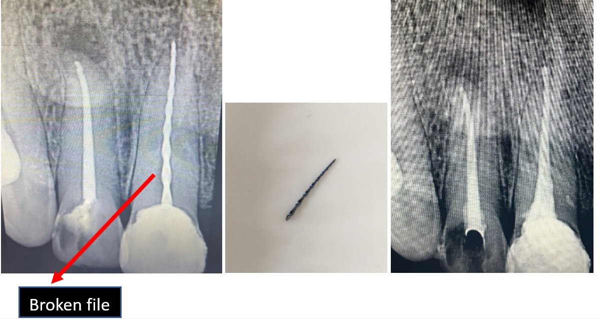
CASE REPORT 2
A 35-year-old male patient presented with persistent pain in the upper left maxillary second molar. The patient reported a history of root canal treatment initiated by a general dentist, which was complicated by a broken file in the mesiobuccal root. Clinical examination revealed tenderness to percussion and palpation in the maxillary second molar region.
A periapical radiograph confirmed the presence of a separated instrument in the mesiobuccal canal of the right maxillary second molar .The diagnosis was symptomatic apical periodontitis associated with a broken file in the mesiobuccal root canal of the right maxillary second molar.
The treatment plan involved the removal of the broken file, complete cleaning and shaping of the root canals, and obturation followed by a definitive coronal restoration.The previous temporary restoration and insufficient access cavity were refined to improve visibility and access to the root canals.
The canals were irrigated with 2.5% sodium hypochlorite (NaOCl) to remove debris. An electronic apex locator (Root ZX II, Morita) was used to determine the working length of the root canals.
Using a high magnification dental operating microscope, the staging platform was created with a Satelec ET20 ultrasonic tip (Satelec Acteon, France) to expose 2-3 mm of the broken file.The Satelec ET25 ultrasonic tip was used to apply ultrasonic energy around the broken file in an anticlockwise direction to loosen it from the canal walls.
- Endodontic micro forceps (Broken Instrument Removal Kit, Zumax, China) were employed to grip and retrieve the broken file from the root canal.The root canals were further prepared using ProTaper Next (Dentsply Maillefer, Switzerland) instruments up to the desired apical size. Irrigation with 2.5% NaOCl and 17% EDTA was performed, followed by activation with an EndoActivator (Dentsply Maillefer, Switzerland).
The root canals were dried with paper points and filled using cold lateral compaction with gutta-percha (ProTaper Next® Gutta Percha, Dentsply Tulsa Dental, Switzerland) and MTA Fillapex sealer.

DISCUSSION
The removal of broken endodontic files is a challenging yet crucial aspect of endodontic treatment. Broken instruments can obstruct the thorough cleaning, shaping, and obturation of the root canal system, potentially leading to treatment failure. In this case, the broken file in the maxillary first molar posed significant challenges due to the complex anatomy of the tooth and the risk of further complications such as perforation or extruding the fragment apically.4
The use of ultrasonic instruments assisted by a dental operating microscope is a conservative and effective approach for removing broken files. Ultrasonics can precisely erode the surrounding dentine without causing significant damage to the root structure or periodontal tissues. However, ultrasonic instruments alone might not always achieve complete file removal, especially if the file is deeply embedded or long. This necessitates the use of additional tools such as microtubes and screw wedges to facilitate retrieval.5
One major concern with the use of ultrasonic instruments is the generation of heat, which can damage periodontal tissues. In this case, EDTA irrigation was employed during the activation of the ultrasonic tips at the lowest power setting to manage the heat and improve the cleanliness of the root canal walls. This approach minimized the risk of thermal damage while ensuring effective removal of the broken file.3
MTA Fillapex sealer was used for obturation due to its beneficial properties, including the ability to stimulate tissue healing, provide excellent sealing, and offer antimicrobial activity. Its high radiopacity aids in confirming the quality of the obturation radiographically. Additionally, MTA Fillapex releases calcium ions, which promote hard tissue formation and enhance the healing process, making it an ideal choice for cases involving periapical lesions.2
The dental operating microscope played a critical role in the successful retrieval of the broken file. Enhanced magnification and illumination allowed for precise manipulation and minimized the risk of procedural errors. The microscope facilitated the creation of a staging platform and the application of ultrasonic energy in a controlled manner, ensuring effective loosening and retrieval of the file.6
The successful removal of the broken file and subsequent obturation led to the resolution of the patient's symptoms. Follow-up radiographs showed a significant reduction in apical radiolucency, indicating successful healing. This case underscores the importance of advanced endodontic techniques and technologies in managing complex cases and achieving favorable outcomes.
CONCLUSION
This case highlights the effectiveness of using ultrasonic instruments, advanced visualization, and appropriate sealing materials in the management of broken files. The conservative approach ensured minimal damage to the tooth structure while achieving complete file removal and successful root canal treatment. Continued advancements in endodontic tools and techniques will further improve the management of such challenging cases, enhancing treatment success and patient outcomes.
REFERENCES
- Shenoy A., Mandava P., Bolla N., Vemuri S. A novel technique for removal of broken instrument from root canal in mandibular second molar. Indian Journal of Dental Research. 2014;25(1):107–110. doi: 10.4103/0970-9290.131157.
- Cohen S., Hargreaves K. Pathway’s of the Pulp. 11th. Kansas, MO, USA: Elsevier; 2016.
- Lambrianidis T. Management of Fractured Endodontic Instruments. Cham, Switzerland: Springer; 2018
- McGuigan M. B., Louca C., Duncan H. F. Clinical decision-making after endodontic instrument fracture. British Dental Journal. 2013;214(8):395–400. doi: 10.1038/sj.bdj.2013.379.
- Gluskin A. H., Ruddle C. J., Zinman E. J. Thermal injury through intraradicular heat transfer using ultrasonic devices: precautions and practical preventive strategies. The Journal of the American Dental Association. 2005;136(9):1286–1293. doi: 10.14219/jada.archive.2005.0346.
- Banerjee S. Bypassing a broken instrument in a severely curved root canal: a case report. Indian Journal of Conservative and Endodontics. 2017;2(3):115–118.
Disclaimer from Guident
The views and opinions expressed in articles published in Guident are solely those of the authors and do not necessarily reflect the views of the editors, the editorial board or the publisher, Ivory India. Guident does not endorse any product, technique or treatment described. Clinical content is intended for qualified dental and medical professionals and is not a substitute for individual clinical judgement. Guident, its editors and the publisher accept no liability for any consequence arising from the use of information published in the journal.
How to cite this article
Dr. Renu Rajput, Dr. Rohit Babu, Dr. Anil Dhingra. Retreiving a Broken File Using Ultrasonic: Two Cases Report. Guident. 2024 Jul;17(8). Available from: https://guident.in/articles/endodontics/retreiving-a-broken-file-using-ultrasonic-two-cases-report.html

