Esthetic Rehabilitation in Anterior Zone - a Case Report
Department of Prosthodontics, Crown & Bridge and Implantology, K.D. Dental College and Hospital, Mathura UP.
INTRODUCTION
The quality of the patient's life is significantly impacted by edentulism. It's crucial to understand that patients receive care that provides them with improved comfort, aesthetics, and dental health. Metal-ceramic prostheses are the gold standard in dentistry, with acceptable aesthetics. Many people nowadays want to improve the appearance of their smile. PFM prosthesis combine the best of both worlds. They are highly aesthetic, as are all ceramic crowns, but far less expensive comparatively. Hence they are extremely popular. Durability is one of their main advantages. They are the strongest and most resistant to fracture of all the aesthetic options for dental prosthesis. Porcelain fused to metal (PFM) prostheses have also been used frequently in implant dentistry when a patient is partially dentate.
This case report describes esthetic rehabilitation of anterior zone using implant supported mandibular prosthesis opposing fixed maxillary prosthesis.
CASE REPORT
A47-year-old female patient reported to the department of Prosthodontics, K.D. Dental College and Hospital, Mathura, complaining of missing mandibular anterior teeth and unaesthetic appearance of maxillary anterior teeth. Patient gave history of getting implants placed 5 years back wherein the prosthesis fractured in one year of it’s cementation. Patient emphasized on replacement of the missing mandibular teeth first and then restoration of the maxillary anterior teeth. Clinical examination Figure 1 revealed missing 31, 32, 41, 42 and 43, faulty splinted prosthesis wrt 11, 12, 13, 21, 22, 23, pink discoloration wrt 21, grade 3 mobility wrt 21, grade 2 mobility wrt 11, spacing betweeen 11 & 21, occlusal caries wrt 35 and generalized gingival recession, generalized plaque and calculus.

Figure 1-Preoperative intraoral view
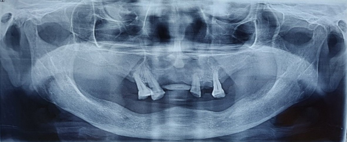
Radiographic examination Figure 2 revealed a mandibular radio-opacity in region of 33 & 42, of previously placed implants and splinted 11, 12, 13, 21, 22 & 23. The Orthopantomogram also revealed generalized interdental bone loss, grossly decayed 27 and periapical radiolucency in relation to 11, 21 & 35.
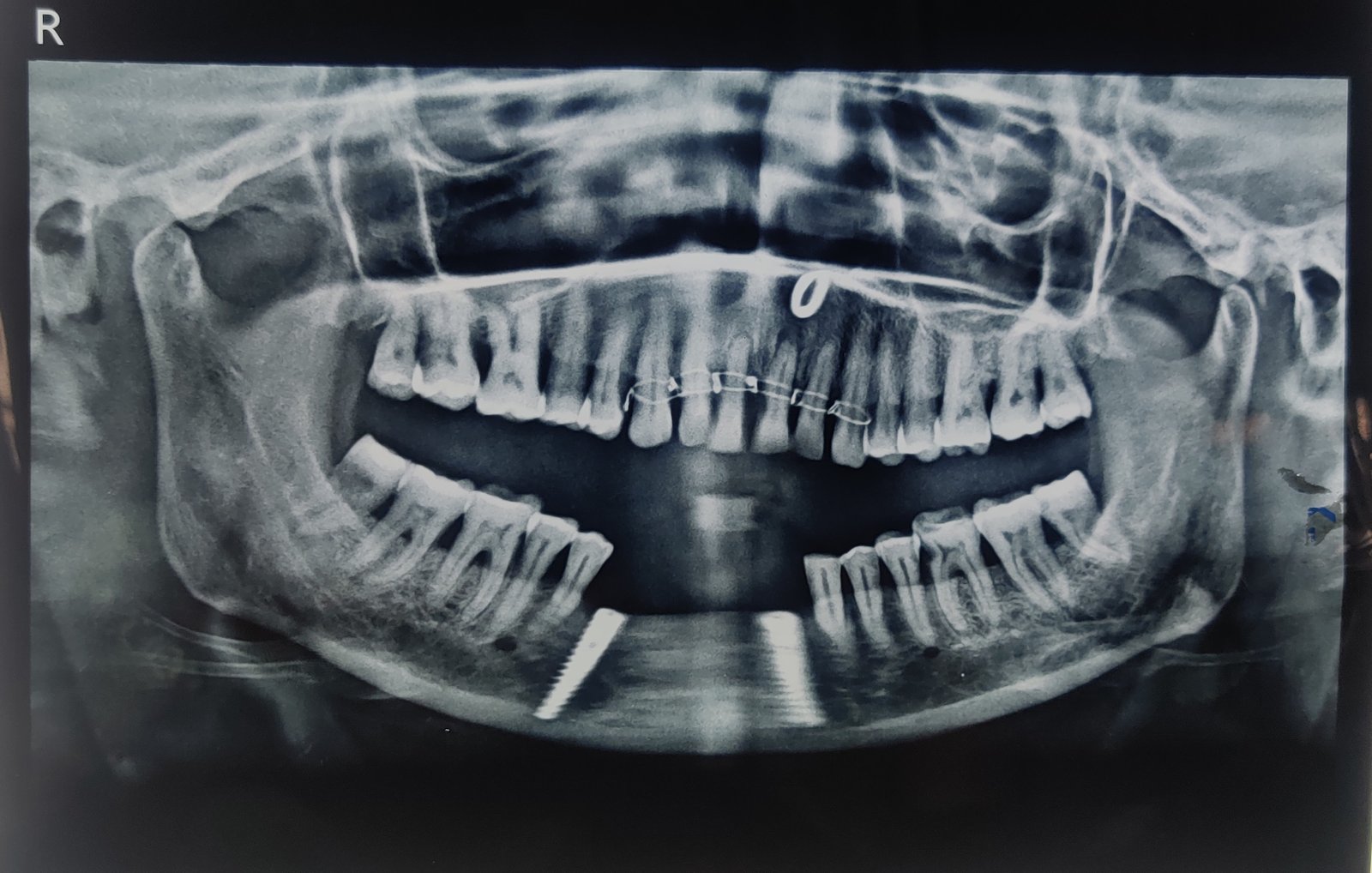
Figure 2- Preoperative orthopantamogram
The patient was diagnosed with the following problems.
- Partially edentulous mandibular arch in relation to previously placed implants in region of 32 & 43
- Faulty prosthesis wrt 11, 12, 13, 21, 22, 23
- Dental caries with chronic apical pulpitis wrt 35.
- Chronic apical pulpitis wrt 11 & 21 and Pink tooth of Mummery wrt 21.
- Chronic generalized periodontitis.
Treatment plan included removal of faulty prosthesis followed by oral prophylaxis. Root canal treatment wrt 11, 21 & 35 and followed by esthetic rehabilitation of maxillary & mandibular anteriors.
The faulty prosthesis wrt 11, 12, 13, 21, 22, 23 was removed followed by oral prophylaxis. Treatment options presented for replacement of missing mandibular teeth was an implant supported cement retained restoration. This treatment option was favourable to the patient. Diagnostic impression of the maxillary and mandibular arches were made using irreversible hydrocolloid.
SURGICAL PHASE
Crestal incision revealed implant positions. [Figure 3, 4]
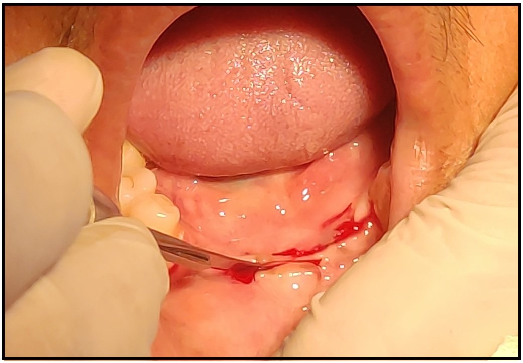
Figure 3

Figure 4
Healing abutments were placed over the implants. [Figure 5] RVG was taken to ensure correct placement of the healing abutments. [Figure 6]
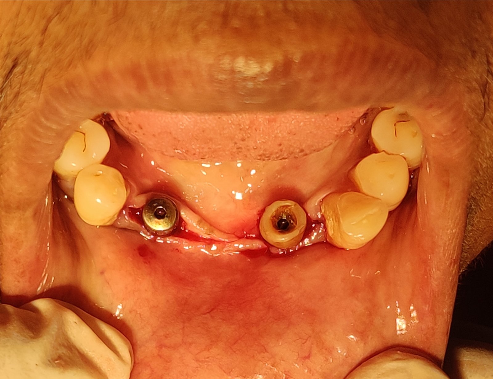
Figure 5

Figure 6
Flaps were approximated and five interrupted sutures were given. [Figure 7] Patient was recalled after a week for removal of the sutures.
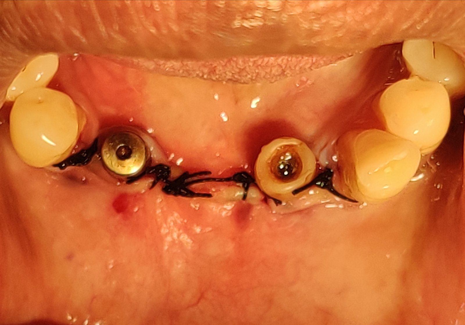
Figure 7
After 3 months, impression copings were placed. [Figure 8] RVG of impression copings was taken. [Figure 9]

Figure 8
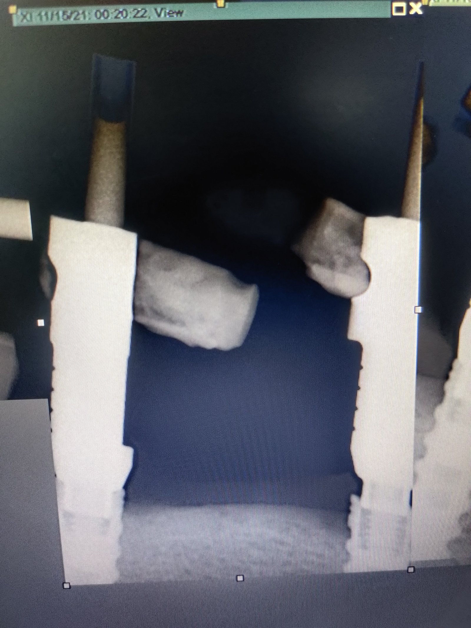
Figure 9
PROSTHETIC PHASE
Copings were splinted and bonded using Flowable composite. Impression tray (plastic) was selected and marked in region of coping orifice for open tray impression. Marked region were relieved in the tray and it was checked in the patient’s mouth. 3M PVS tray adhesive was applied onto the tray. Open tray impression was made of the mandibular arch using PVS soft putty & PVS light body. Gingitech highly elastic PVS was used for gingival mask. [Figure 10] Alginate impression was made of the maxillary arch.


Figure 10
Boxing of the Mandibular impression was done. Die stone was poured as first layer and the base was built using Dental stone. Gingival mask from the impression was carefully placed in it’s counter part in the cast obtained. Open tray transfer copings were tried in the cast [Figure 11]
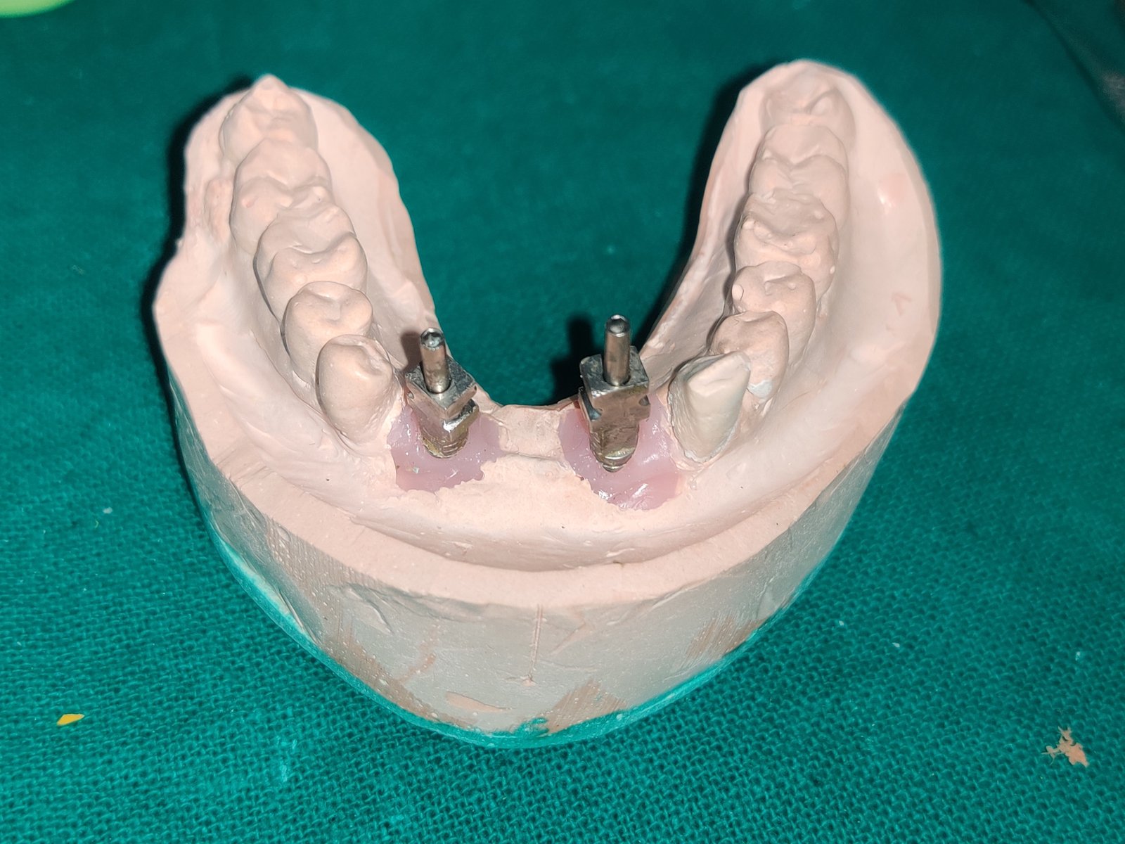
Figure 11
Jig was prepared by splinting the transfer copings using thread & Pattern resin (GC) over it.
Jig trial was done in the patient & RVG was taken. [Figure 12, 13]

Figure 12
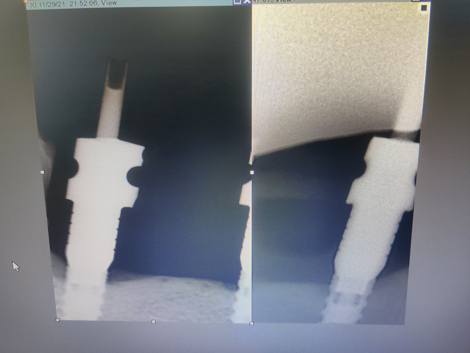
Figure 13
Angulated abutments were placed and checked via RVG. [Figure 14, 15]

Figure 14

Figure 15
Metal coping trial was done. [Figure 16, 17]

Figure 16
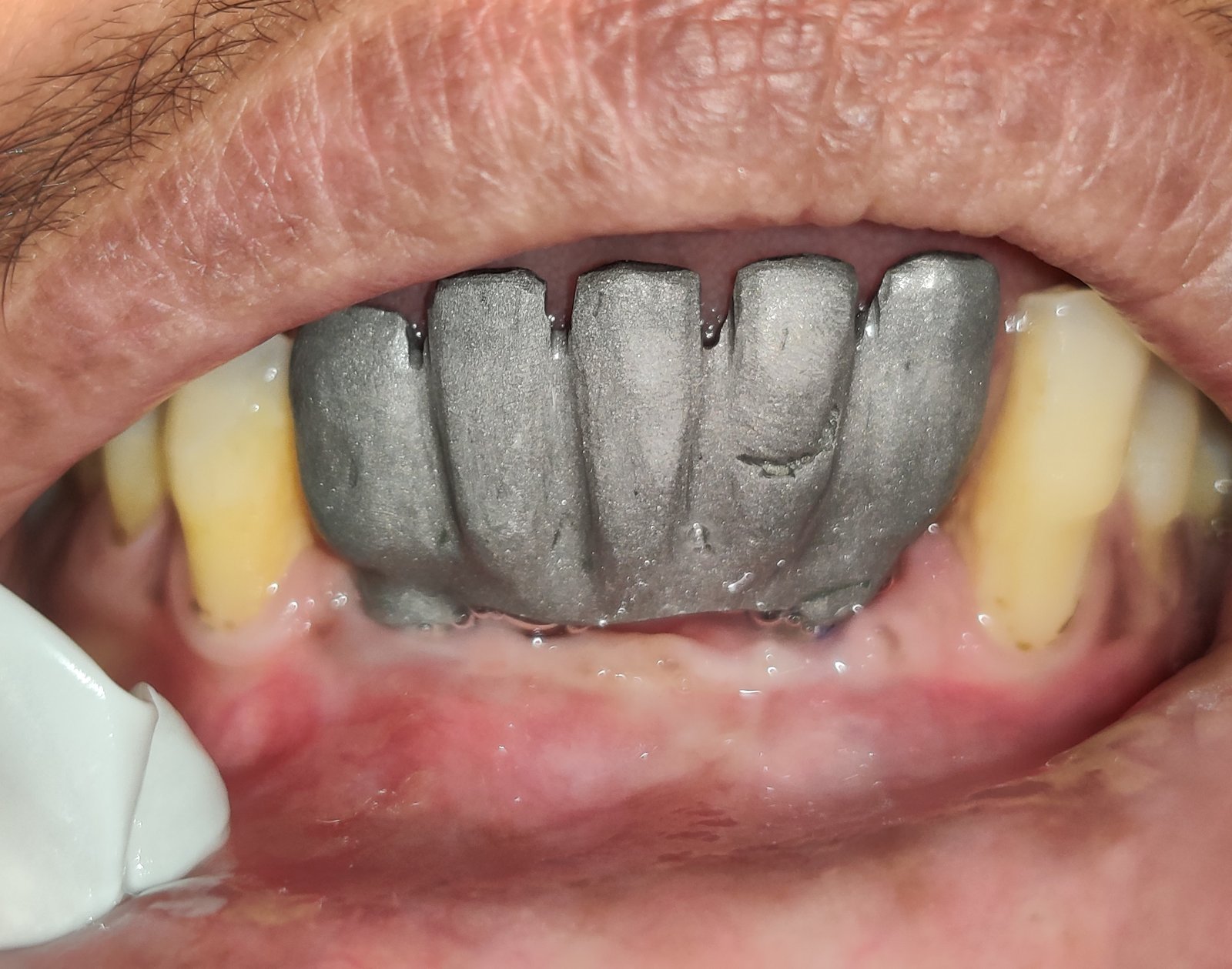
Figure 17
A-3.5 shade was selected for PFM restoration. Prosthesis was fabricated and cemented wrt 31, 32, 41, 42 & 43. For this particular patient’s treatment, pink ceramic was used for the greater ridge resorption anteriorly. [Figure 18, 19]
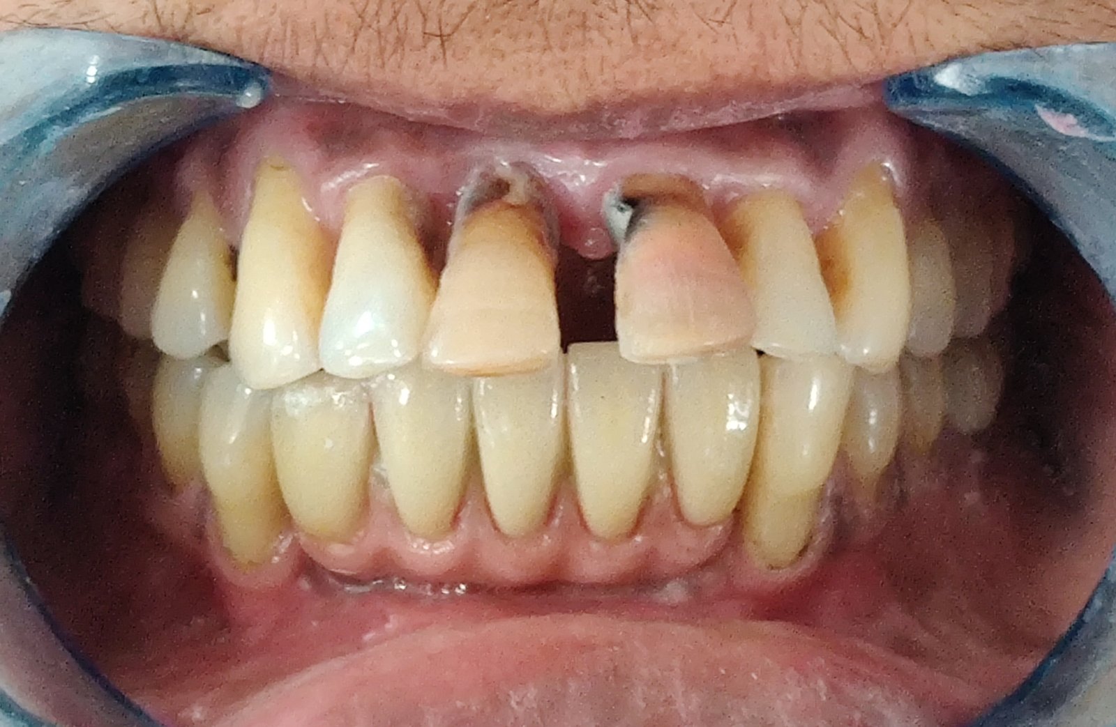
Figure 18

Figure 19
Patient was satisfied with the replaced missing mandibular anteriors and now was willing for treatment of maxillary teeth. The following teeth were extracted; 11 & 21. A tooth supported PFM fixed partial denture was planned for replacement of the missing maxillary anterior teeth using 12, 13, 22 & 23 as abutments. [Figure 20]
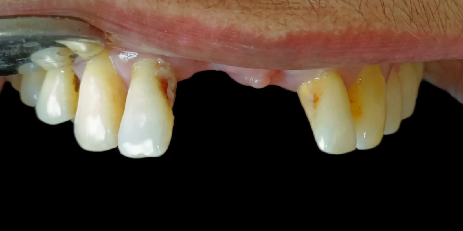
Figure 20
Due to greater proclination of anterior teeth intentional RCT was done wrt 12, 13, 22 & 23. Tooth preparation was done wrt 12, 13, 22 & 23 to receive PFM fixed partial prosthesis. [Figure 21] Alginate impression was made and cast was poured using dental stone for making provisional restoration.
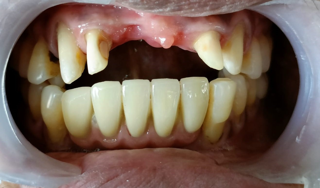
Figure 21
Final impression was made using 3M PVS soft putty & 3M PVS light body impression material. [Figure 22]
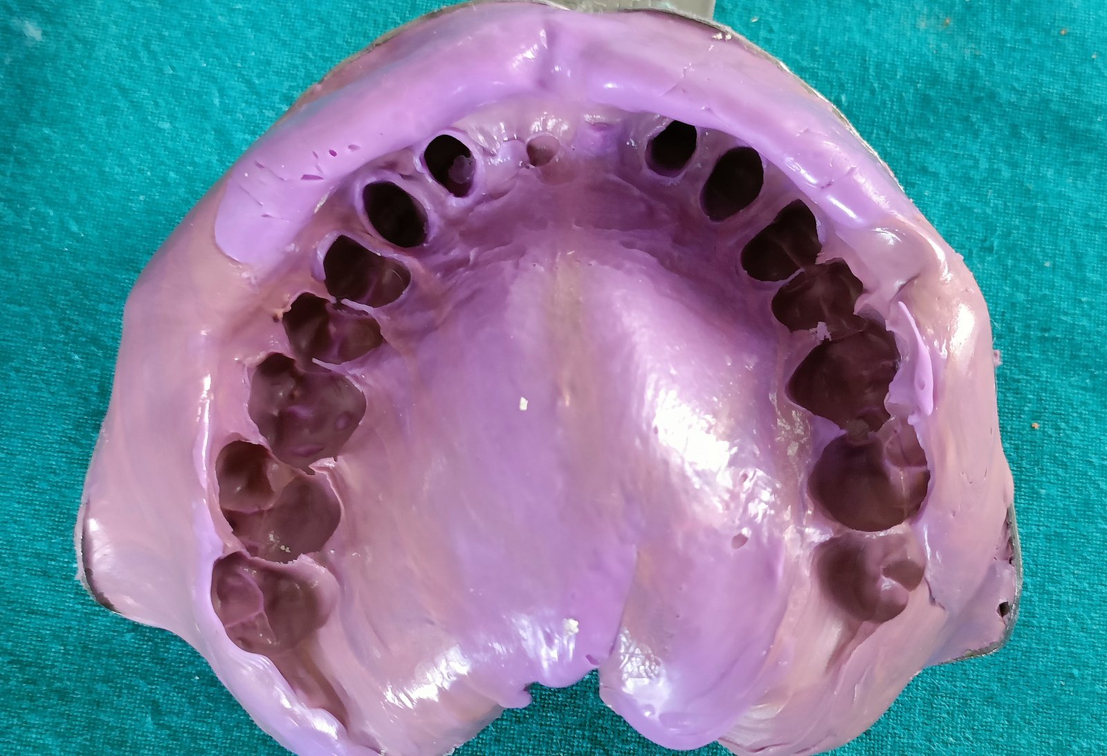
Figure 22
After a week, the provisional prosthesis was removed and final PFM fixed prosthesis was cemented with 12, 13, 22 & 23 as abutments. [Figure 23]

Figure 23
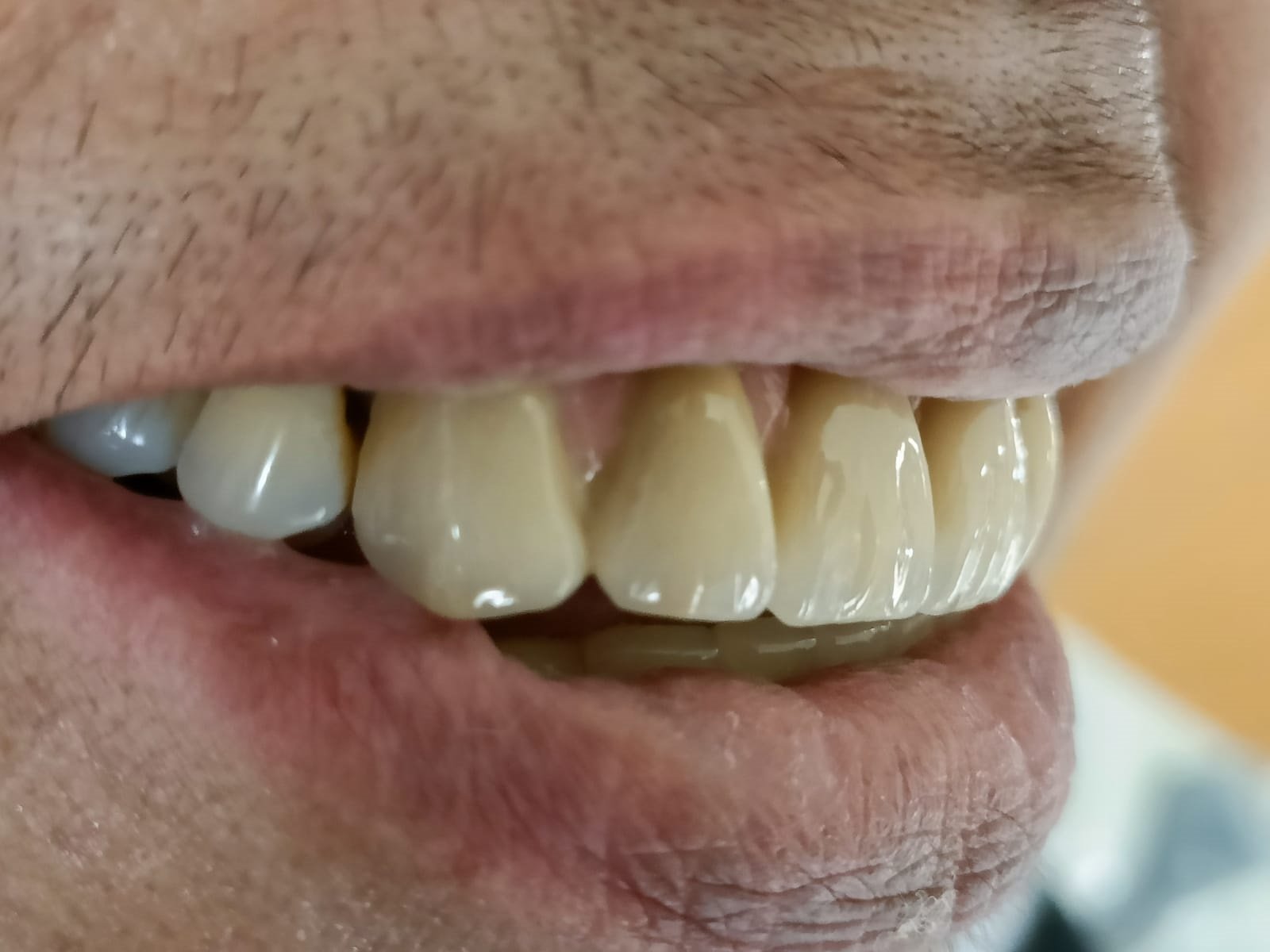
Figure 24

Figure 25

Figure 26
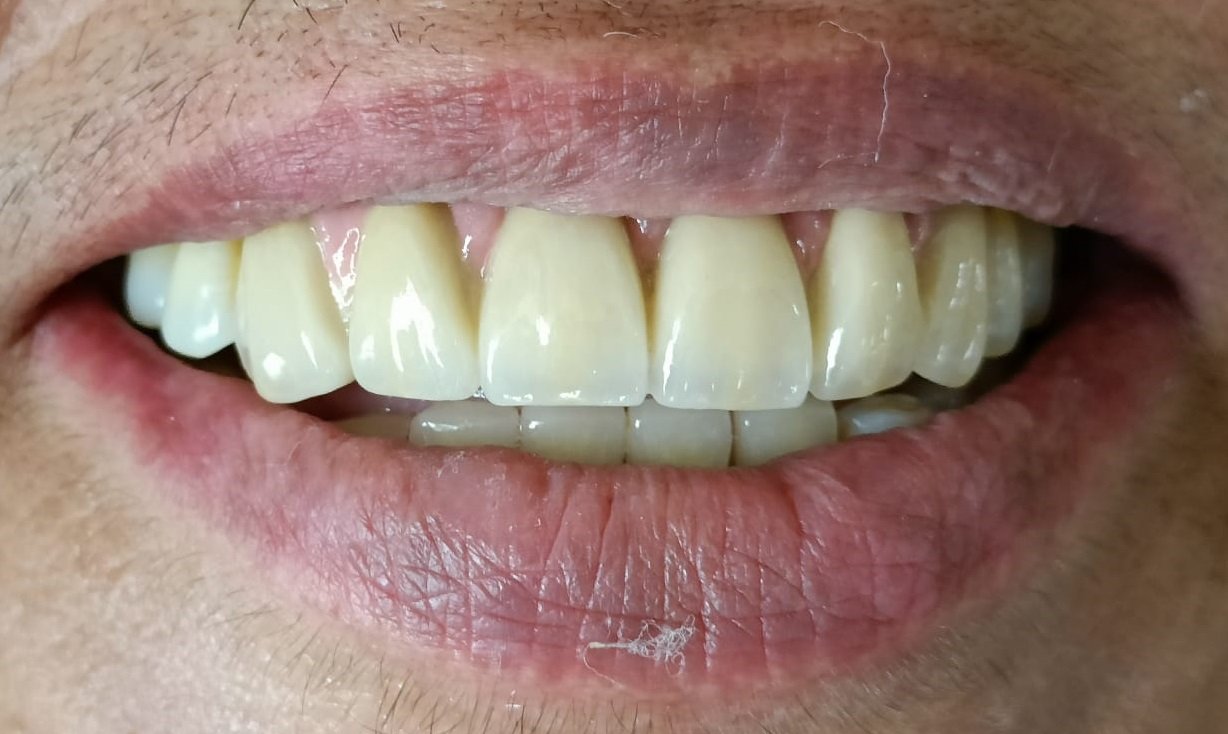
Figure 27
The patient was immensely satisfied with her smile. [Figure 24, 25, 26, 27]
CONCLUSION
In order to treat the maxillary and mandibular long span partially edentulous jaw, this article discusses the clinical procedures for the aesthetic and functional rehabilitation using fixed tooth supported prosthesis in maxillary anterior opposing an implant supported prosthesis in mandibular anterior region. It may, therefore, be concluded that this treatment enhanced the overall oral health & smile of the patient.
Disclaimer from Guident
The views and opinions expressed in articles published in Guident are solely those of the authors and do not necessarily reflect the views of the editors, the editorial board or the publisher, Ivory India. Guident does not endorse any product, technique or treatment described. Clinical content is intended for qualified dental and medical professionals and is not a substitute for individual clinical judgement. Guident, its editors and the publisher accept no liability for any consequence arising from the use of information published in the journal.
How to cite this article
Dr. Mehr Yazdani, Dr. Neha Srivastava, Dr. Rahul Nagrath. Esthetic Rehabilitation in Anterior Zone - a Case Report. Guident. 2022 Jun;15(7-8). Available from: https://guident.in/articles/prosthodontics/esthetic-rehabilitation-in-anterior-zone-a-case-report.html