Seal the Split-a Case Series
Department of Conservative Dentistry and Endodontics, Seema Dental College And Hospital, Rishikesh
INTRODUCTION
A successful endodontic treatment requires clinician sound knowledge on root canal morphology, cleaning, shaping and obturation of the teeth. Mandibular premolars have complex anatomies withhigher incidence of two canals in mandibular first premolars was reported in several populations, upto 50% in Indian populations, 40% in Middle Eastern populations from Kuwait, Caucasian populations had a variable incidence of two canals in about 10.7–36% The incidence of three or more canals in mandibular first premolars was considerably lower (0–5%). The prevalence of three root canals with three distinct orifices that end in separate foramina was reported to be 0.4% by Zillich and Dowson in 1973. Vertucci et al. reported that the mandibular second premolar had one root canal at the apex in 97.5% and two canals in only 2.5% of the teeth; however, three root canals were scarce Factors that are related to the variations found in premolars include ethnicity, gender, age.
The aim of this article is to evaluate anatomical variations of mandibular premolar teeth.
CASE REPORT-1
A 50 year old male patient came to the department of conservative dentistry and endodontics with the chief complaint of pain in lower left back tooth region for 5 days. Medical history was non contributory. Clinical examination showed deep proximal caries and radiographic examination showed proximal caries involving enamel dentin and pulp with a bifurcated root at apical 3rd. CBCT revealed splitting of canal at the level of apical 3rd. Cold test gave a lingered response and electric pulp tester reading suggested symptomatic irreversible pulpitis.

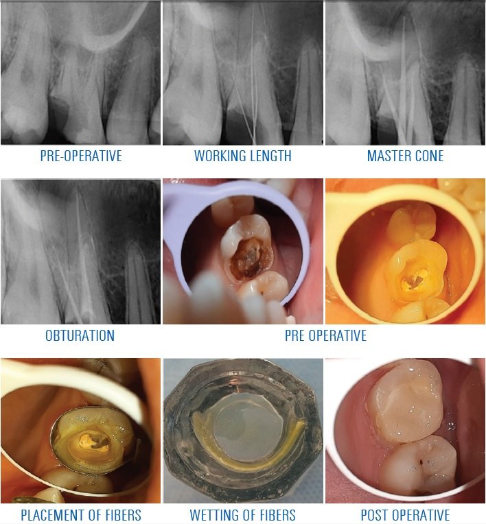
Nonsurgical endodontic treatment was planned i.r.t 44. After the administration of the local anesthetic agent (2% lidocaine with 1: 100,000 epinephrine) access was initiated and a Working length was established with the use of an apex locator and confirmed by a radiograph. Cleaning and shaping was done up to the master apical file using 2% Naocl and 17% EDTA. After final rinse with normal saline, canals were dried and obturated with gutta-percha and MTA fill apex as a sealer using downpack and backfill method.

CASE REPORT-2
A 22 year old female patient came with the chief complaint of pain in lower left back tooth region since 10 days. Patient gave dental history of root canal treatment in the same tooth 2 years back. On radiographic examination, there was periapical radioleucency involving 35. On the basis of clinical and radiographic findings, a diagnosis of apical periodontitis was made w.r.t. 35. Treatment plan decided was non surgical retreatment i.r.t 35. A written consent was taken from the patient after explaining the treatment protocol. In the first visit, the old guttapercha was removed using H files and was checked radiographically. Intra canal medicament was placed and the patient was recalled after 15 days. In the second appointment, first two canals were obturated, then the GP was shredded till the deep split and then the third canal was obturated, which was missed by the last dentist. Final post endodontic restoration was done using composite in the third appointment.

DISCUSSION
Mandibular premolars have the most anatomical variation and are a challenge to the clinician.
Radiographic analysis with SLOB technique and CBCT are essential in providing insight into the number of existing root canals. Sudden change in radiographic density and sudden narrowing of root canal space usually indicates an additional canal. Along with radiographic analysis the use of loupes, anatomical landmarks in pulp chamber, use of sodium hypochlorite for champagne bubble test and dyes may be helpful in locating additional canals.
In our first case report a preoperative radiograph with sudden disappearance of root canal suggested of an extra canal. CBCT analysis confirmed the presence of extra canal. Careful observation and inspection of the pulpal floor and also pulpal wall is mandatory to avoid the unexpected missing of orifices that may cause unsuccessful endodontic treatment. Colour changes in pulpal floor also helps in locating canal Access was determined and gates gladden drill was used to enlarge the orifice. Only after using gates gladden drill the second orifice was found. Microscopes and loupes are commonly used to explore the pulp chamber in order to find orifices. Advantages of using a microscope and loupes for conventional endodontics include better visualization of the pulp chamber floor and walls that prevents inadvertent missing of orifices. Cleaning and shaping was done with rotary files and canals were obturated with down pack and backfill.
In our second case report the radiographic examination showed a periapical radiolucency suggesting of failed root canal treatment due to a missed canal.
After removing the gutta percha the 3rd canal was found and a working length was determined with k file. After cleaning and shaping of the root canals the teeth was oburated with downpack and were restored with composite.
It is assumed that careful observation and inspection of the pulpal floor and also pulpal wall with innovations in diagnosis, magnification, operative instruments and techniques is mandatory to avoid the unexpected missing of orifices that may cause unsuccessful endodontic treatment.
CONCLUSION
Successful and predictable endodontic treatment requires knowledge of normal anatomy and variations. In the case where radiographic images are not helpful to clarify root canal anatomy and aberrations, magnification devices are recommended. Using dye for enhancement of color contrast is helpful in visualizing deeply situation orifices and variations.
REFERENCES
- Mehrdad Lotfi, Sepideh Vosoughhosseini A mandibular second premolar with three canals and atypical orificesJournal of Oral Science, Vol. 50, No. 3, 363-366, 2008
- Denzil Albuquerque, Jojo Kottoor Endodontic and Clinical Considerations in the Management of Variable Anatomy in Mandibular Premolars: A Literature Review Volume 2014, Article ID 512574, 11 pages
- Deena Jassem alenezi, Saad A AL Nazhan root and Canal Morphology of Mandibular Premolar Teeth in a Kuwaiti Subpopulation: A CBCT Clinical StudyEur Endod J 2020; 3:248-56
- Blaine M. Cleghorn,William H. Christie The Root and Root Canal Morphology of the Human Mandibular First Premolar: A Literature Review JOE — Volume 33, Number 5, May 2007
- Zahra Borna, Saeed Rahimi Mandibular Second Premolars with Three Root
- Canals: A Review and 3 Case ReportsIranian Endodontic Journal 2011;6(4):179-182
Disclaimer from Guident
The views and opinions expressed in articles published in Guident are solely those of the authors and do not necessarily reflect the views of the editors, the editorial board or the publisher, Ivory India. Guident does not endorse any product, technique or treatment described. Clinical content is intended for qualified dental and medical professionals and is not a substitute for individual clinical judgement. Guident, its editors and the publisher accept no liability for any consequence arising from the use of information published in the journal.
How to cite this article
Dr. Sugandha Sati, Dr. Aryama Balodi, Dr. Anil Dhingra. Seal the Split-a Case Series. Guident. 2022 Jun;15(7-8). Available from: https://guident.in/articles/endodontics/seal-the-split-a-case-series.html