
Surgical Correction of Skeletal Class II
A 25 year old femalereported to the department with the chief complaint of deviated mandible to the left side. She gave history of trauma due to fall when she was 5 months old and underwent surgery for the same. Patient had undergone orthodontic treatment at 12 years of age with the extraction of all first premolars.
CLINICAL EXAMINATION
On Intra oral examination, all permanent teeth were present except the 4 premolars. The maxillo-mandibular relationship can be summarized with both the Molar and Canine relationship being Class II bilaterally. The Incisor relationship was Class II Div 1 pattern with 13 mm of overjet and 6.5 mm of overbite. Crossbite was seen in the right segment of the arch in relation to 45, 46 and 47. The lower midline was shifted to left by 7 mm.

On Functional analysis, the mandibular midline shift was evident in habitual occlusion and in rest position. The maximum interincisal distance was 33 mm. there were no deviations in lateral and frontal path of opening and closure.
RADIOGRAPHIC EXAMINATION
Pretreatment Orthopantomograph (OPG), PA view and Lateral Cephalogram were made. Various cephalometric analysis were done including Steiner,s, Downs, Tweeds, McNamara,s and others. The patient had Class II skeletal apical base with orthognathic maxilla (SNA=82o) and retrognathic mandible (SNB=71 o) ; WITS appraisal of +11 mm ; average growth pattern ; reduced interincisal angle (116 o) ; increased upper facial height ; decreased lower facial height ; reduced body length of mandible.


DIAGNOSTIC SUMMARY
Skeletal Class II malocclusion due to retrognathic mandible with reduced mandibular body length and chin length ; Angle’s Class II Molar and Canine relationship bilaterally ; overjet of 13 mm and overbite of 6.5 mm ; crossbite in the right segment of the arch in relation to 45, 46 and 47 ; shift in the lower midline by 7 mm were observed.
RELEVANT CEPHALOMETRIC VALUES

DIAGNOSIS
- Facial asymmetry
- Class II skeletal base due to retrognathic mandible
- Scissor bite on right side
- Decreased mandibular body length
- Recessive Chin
- Occlusal cant to left side
- Increased upper facial height
- Angle’s Class II Molar and Canine relationship
- Maxillary anterior proclination and mandibular anterior crowding
- Convex profile
- Everted lower lip
TREATMENT PLAN
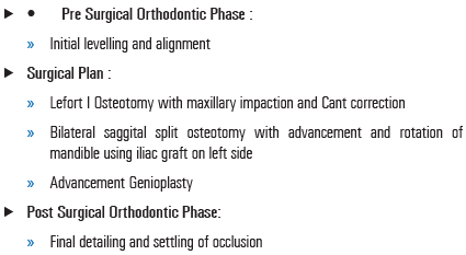
PRE SURGICAL PHOTOGRAPHS :

POST SURGICAL PHOTOGRAPHS :


POST TREATMENT INTRAORAL

POST TREATMENT EXTRAORAL:

Disclaimer from Guident
The views and opinions expressed in articles published in Guident are solely those of the authors and do not necessarily reflect the views of the editors, the editorial board or the publisher, Ivory India. Guident does not endorse any product, technique or treatment described. Clinical content is intended for qualified dental and medical professionals and is not a substitute for individual clinical judgement. Guident, its editors and the publisher accept no liability for any consequence arising from the use of information published in the journal.
How to cite this article
Dr. Suraj Prasad Sinha, Dr. U.S. Krishna Nayak Department of Orthodontics and Denofacial Orthopedics. Surgical Correction of Skeletal Class II. Guident. 2016 Dec;10(1). Available from: https://guident.in/articles/orthodontics/surgical-correction-of-skeletal-class-ii.html


