Buccal Fat Pad for Closure of Intra-Oral Defects
Abstract
The pedicled buccal fat pad has been widely used for the repair of intra-oral defects. It is locally available, can be easily harvested with minimal donor site morbidity. This
article presents a few cases of intra-oral defects which were reconstructed utilizing the buccal pad of fat.
Key words: Intra-oral defects, buccal fat pad.
INTRODUCTION
The buccal fat pad (BFP) is a mass of specialized fatty tissue which is distinct from subcutaneous fat. It was first recognized as such, and described by Bichat in 1802.3 It had a limited clinical importance for many years and was usually considered a surgical nuisance in the pterygomaxillary space or after injuries of the maxillofacial region. Egyedi (1977)4 was the first to report on the application of the BFP as a pedicled graft for the closure of oroantral and oronasal communications. However its use has been extended for reconstruction of surgical defects of the oral soft and hard tissues.
The anatomy of the BFP is complex. The main mass occupies the buccal space bounded medially by the buccinator muscle and laterally by the masseter muscle, and rests on the periosteum that covers the posterior and buccal aspect of the maxilla. The BFP has a central body with four extensions (Fig 1).
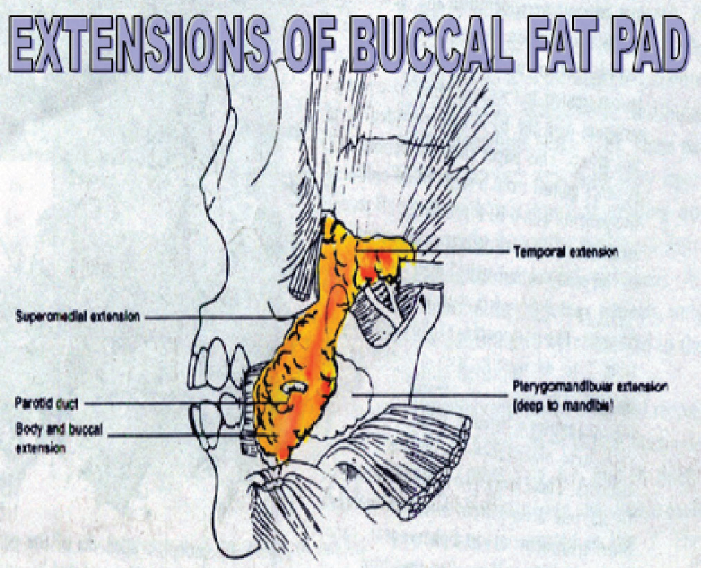
The main body lies above the parotid duct on the anterior border of the masseter muscle and extends deeply to lie on the posterior maxilla. The buccal extension is the most superficial and enters the cheek below the parotid duct. It descends to the mandibular vestibular region and overlies the main part of the buccinator muscle. The pterygoid extension passes down and back to lie on the lateral surfaces of the pterygoid plates. The temporal extension passes upwards below the zygomatic arch, and comprises a deep and superficial portion.
The BFP has blood supply through the branches of the superficial temporal artery, the internal maxillary artery and small branches of the facial artery along with veins by an abundant net of vascular anastomosis. Care is to be taken to avoid severing the supporting vascular plexus and the thin capsule covering the BFP. On an average its volume is 9.6 CC (range 8.3 – 11.9 cc). The average weight was found to be 9.3 gm. The pedicled BFP can cover defects of the posterior maxillae as far as the region of the canine tooth, the hard palate, the soft palate and the retromolar and pterygomandibular regions. Defects up to 3 x 5 cm. can be closed alone with a BFP without compromising the blood supply.
CASE 1
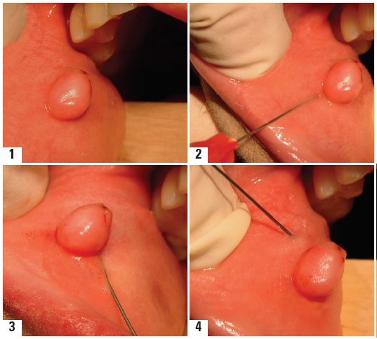
Eight cases of oroantral fistulae were closed by BFP where regional flaps were inadequate to cover up the defects (Fig 2).Infection in the maxillary sinus was brought under control by antral lavage with normal saline via the fistula at 48 hrs interval, until a clear return was obtained.

Fig 1
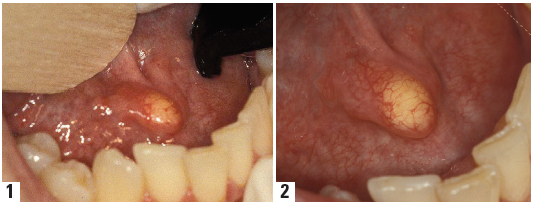
After excision of the fistulous tract with a 3 mm margin divergent incisions were made from each end of the defect. A 1 cm long vertical incision over the reflected periosteum posterior to the zygomatic buttress was given and the BFP was gently teased out, advanced over the bony defect and secured to the palatal mucosa without tension by sutures. The mucoperiosteal flap was replaced in its original position, and sutures inserted between the BFP and the buccal flap. A part of the fat remained exposed in the oral cavity. Post-operative healing was uneventful and sutures were removed after 10 days. On 2nd day the orally exposed fat became yellowish white, on 7th day it became red, on 14th day it changed into bright

Fig 2
pink granulation tissue while on 21st day it turned to be pale pink. Shinkage of the BFP was seen during the healing process. After healing was completed(Fig.3). There was no difference in the level or colour to the local mucosa.
CASE 2
A 42 years old man reported with a white cheek lesion (3cm x 2cm) involving the left buccal mucosa in the molar region which was found to be verrucous caricoma on incisional biopsy. Excision of the white lesion including adequate amount of healthy tissue at the margins was carried out. Through the created surgical defect, an incision was given over the fibres of the buccinator muscle and by blunt dissection with a curved haemostat the fascial

Fif 3
envelope of the BFP was opened and further manipulation mobilized it over the defect easily without undue tension and suturing done (Fig 4) Epithelialisation was almost complete by about 4th week postoperatively.
CASE 3
A 56 years old female patient with oral submucous fibrosis having clinically palpable fibrotic bands on the buccal mucosa unilaterally and partial trismus was treated by excision of the fibrotic band at the level of the occlusal plane away from the Stenson’s onfice and extended posteriorly to the pterygomandibular raphe and extended as for as the first premolar region. The exposed would was further manipulated by blunt dissection to permit full mouth opening without tension. The BFP was mobilized by separating the fibres of buccinator and teasing it out through the wound in sufficient amount so as to cover up the exposed defect without undue tension (Fig 5 A,B ) .Epithelialisation was almost complete in 3-4 weeks. The range of mouth opening measured was 28-34 mm.

Fig 5A

Fig 5B
Case 4
Six cases of medium-sized post surgical defects were reconstructed by the pedicled BFP grafts. All the defects were in the maxilla in the posterior molar regions, created, following surgical removal of cysts or benign tumours. The dead spaces within the bony defects were obliterated by gently packing them with Whitehead Varnish ribbon gauze. A periosteal cut over the reflected periosteum on the undersurface of the mucoperiosteal flap just posterior to the zygomatic buttress was given and the BFP was teased out gently until a sufficient amount was obtained to cover up the defect without tension, care being taken not to rupture the capsule with its rich vascular plexus (Fig 6). The flap was replaced in its original position, and sutures inserted between the fat and the buccal flap so that a part of the fat remain exposed in the oral cavity. No surgical splint or dressings were used.

Fig 6
DISCUSSION
The BFP as a pedicled graft with an axial pattern possesses favourable characteristics for the reconstruction of intra-oral defects. Tideman et al (1986)4 postulated that defects up to 3 x 5 cm. can be closed with BFP alone without compromising the blood supply. However Rapidis et al (2000)2 found that in surgical defects measuring more than 4 x 4 x 3cm. the likelihood of partial dehiscence of the flap is high. However no dehisence was observed in our case though the maximum size was 4x3x4 cm. It might be due to the limited number of cases and a follow up study with a larger number of sample size is required. The procedure is technically easy and the donor site is in close proximity to the defect. It can also be used in conjunction with other local flaps or regional myofascial flaps. There is wide applicability in most cases with minimal jncidence of failure when properly performed.Stajcic (1988)1suggested that the BFP technique should not be regarded as a mere substitution for the buccal sliding flap for repair of orantral fistulae because it is especially indicated in cases with damage to the region of the alveolar buccal or palatal mucoperiosteum. Hanazawa et al (1995)4 further overviewed that the BFP should be used in cases that have either failed with other methods or in patients who need early preparation of a prosthesis. All the eight cases of OAF in this study were chosen where regional flaps were inadequate to cover up the defect.
CONCLUSION
BFP is a readily available tissue for the closure of small to medium sized intra-oral defects and being locally available it is easy to harvest with no donor site morbidity. It is a simple procedure with a minimal incidence of failure and good success rate.
References
- Hanazawa et al : Closure of Oroantal communications using a pedicled buccal fat pad graft. J Oral Maxillofacial Surgery 53 : 771-775, 1995.
- Rapidis DA : The use of the Buccal Fat pad for reconstruction of oral defects : Review of the Literature and Report of 15 cases. J of Oral & Maxillofacial Surgery : 54 : 158-163 = 2000.
- Samman. N et al : The buccal fat pad in Oral reconstruction. Int. J. Oral Maxillofacial Surgery – 1993 : 22: 2-6.
- Yeh C.Y : Application of the buccal fat pad to the surgical treatment of Oral submucous fibrosis. Int. J. Oral Maxillofacial Surgery. 1996 : 25 : 130-133.
- Poeschl PW, Baumann A, Russmueller G, Poeschl E, Klug C,Ewers R. Closure of oroantral communications with Bichat’s buccal fat pad. J Oral Maxillofac Surg 2009;67:1460-6.
- Adeyemo WL, Ogunlewe MO, Ladeinde AL, James O. Closureof oro-antral fistula with pedicled buccal fat pad. A case reportand review of literature. Afr J Oral Health 2004;1:42-6.
- Marzano UG. Lorenz Heister’s “molar gland.” Plast ReconstrSurg 2005;115:1389-93.
- Baumann A, Ewers R. Application of the buccal fat pad in oral reconstruction. J Oral Maxillofac Surg 2000;58:389-92.
- Rapidis AD, Alexandridis CA, Eleftberiadis E, Angelopoulos AP. The use of the buccal fat pad for Sreconstruction of oral defects: review of the literature and report of 15 cases. J Oral Maxillofac Surg 2000;58:158-63.
- Tideman H. Bosdnquet A, Scott J. Use of the buccal fat pad as a pedicled graft. J Oral Maxillofac Surg 1986;44:435-40.
- Hao SP. Reconstruction of oral defects with the pedicled buccal fat pad flap. Otolaryngol Head Neck Surg 2000;122:863-7.
- Dean A, Alamillos F, Garcia-Lopez, A, Sanchez J, Penalba M. The buccal fat pad in oral reconstruction. Head Neck 2001;23:383-8.
- Stuzin JM, Wagstrom L, Kawamoto HK, Baker TJ, Wolfe SA. The anatomy and clinical applications of the buccal pad of fat. Plast Reconstr Surg 1990;85:29-37.
- Dubin B, Jackson IT, Halim A, Triplett WW, Ferreira M. Anatomy of the buccal fat pad and its clinical significance. Plast Reconstr Surg 1989;83:257-62.
- Zhang HM, Yan YP, Qi KM, Wang JQ, Lui ZF. Anatomical structure of the buccal fat pad and its clinical adaptations. Plast Reconstr Surg 2002;109:2509-20.
Disclaimer from Guident
The views and opinions expressed in articles published in Guident are solely those of the authors and do not necessarily reflect the views of the editors, the editorial board or the publisher, Ivory India. Guident does not endorse any product, technique or treatment described. Clinical content is intended for qualified dental and medical professionals and is not a substitute for individual clinical judgement. Guident, its editors and the publisher accept no liability for any consequence arising from the use of information published in the journal.
How to cite this article
Dr. Asish Kumar Das, Dr. Ranendra Nath Poddar Dr. Vikash Jha. Buccal Fat Pad for Closure of Intra-Oral Defects. Guident. 2013 Feb;6(4). Available from: https://guident.in/articles/oral-surgery/buccal-fat-pad-for-closure-of-intra-oral-defects.html