Bilateral Nasolabial Cyst- a Rare Case Report of Nonodontogenic Cyst
K.D Dental college & Hospital, Mathura, India
ABSTRACT
The nasolabial cyst is an uncommon non-odontogenic cyst arising in the maxillofacial tissues. Nasolabial cyst, also known as Klestadt's cyst is an uncommon nonodontogenic cyst. Bilateral nasolabial cysts are rarer and less than 10 cases have been reported in the literature. This lesion presents in an extraosseous location in the region of the nasolabial fold and can cause swelling in the furrow, alar nose elevation, and upper lip projection. Despite the uncommon occurrence of nasolabial cysts, it is important to recognize the characteristics of this lesion.
Key words: Cyst,nasolabial, non-odontogenic.
INTRODUCTION
The nasolabial cyst (NC) is a rare non-odontogenic cyst originating in the maxillofacial soft tissue1. It representsabout 0.7% of all cysts in the maxillofacialregion, 2.5% of the non-odontogenic cysts. These cysts unless infected causepainless swelling around the nasal vestibule and upper lip and infrequently lead to nasal stuffiness.2Typically, theyappear as a swelling at canine fossa, upper lip, gingivo-labial sulcus, nasal alae and nasal vestibule. Despite the fact thatthey are soft tissue cysts situated extra‑osseously and sometimes cause bone destruction. The initialdiagnosis and treatment is usually made in early stages because the lesion causes cosmetic problems; very rarely it becomes large in dimensions. Commonly seen in adults, it has peak prevalence in the 4th‑5th decade of life.3 Agreater incidence is seen in females (4:1). It is usually unilateral in occurrence with no predilection in side. However, 11.2% cases have been reported to be bilateral.Although uncommon in occurrence, it is imperative for the clinician to make an accurate diagnosis and provide appropriate treatment.8
CASE REPORT
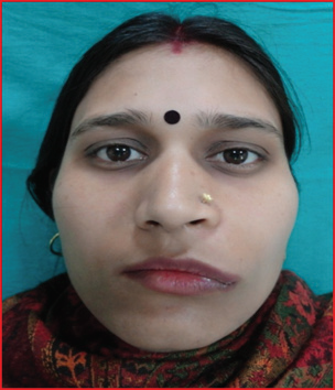
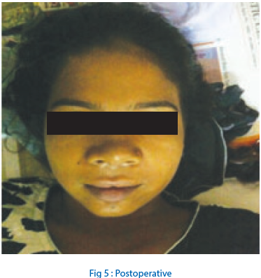
A 24 year old female patient came to the department of oral & maxillofacial surgery for the assessment of bilateral mass in the region of ala of the nose.(Fig 1)The patient was unable to describe precisely how long the lesion had been present. The lesion was painless and there was no history of epistaxis or nasal dischargeExtraoral examination revealed facial asymmetry with slight elevation of the alanasi and deformation of the left nasolabial sulcus.(Fig 1) On palpation, the lesion was nodular, soft, fluctuant, well circumscribed bilaterally and did not extend beyond the soft tissues.A fullthickness incision was reflected along the crest of maxillary alveolar ridge from canine to canine regionbilaterally(Fig2,3) and blunt dissection was done by periosteal elevator(Fig 4)&exposed two smooth, wellcircumscribed cystic swellings just below the nasal floor. There had been scooping of the anterior maxilla on the left side due to expansion of the cyst. In contrast, morphologyof alveolar bone on right side was not affected.Suturing done with 3-0vicryl. No postoperative complications occurred.(Fig 5)
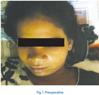

Fig 2: Intraoperative right cystic swelling
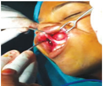
Fig 3: Intraoperatively left cystic
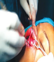
Fig 4: Blunt dissection by periosteal elevator swelling
DISCUSSION
Nasolabial cysts were first described by Zuchercandl in 1892 . They are nonodontogenic masses that can be seen in the maxillofacial area. In the literature, the lesions are name as nasolabial cyst, nasoalveolar cyst and Klesdath tumour.4The lesion is submucosal and extraosseous, it expands via the gingivobuccal sulcus and expands all the soft-tissues outwards. Usually the cysts are seen in the 4th-5th decade of lifetime. The incidence of bilateral cyst is 10% in the literature. There are three theories for the formation of the cyst-
(1) The cyst is formed embryologically by detention cells in the maxilla, medial. and lateral nasal wall.
(2) The cyst is formed embryologically by detention cells from the inferior nasolacrimal channel redundant cells.
(3) The cyst is formed embryologically by detention cells from the inferior nasolacrimal channel endodermal cells .
Exposure to trauma accelerates the formationof the cyst. The differential diagnosis of the cyst must be made with central line cysts, cyst of maxilla, odontogenic cysts, periapical cysts, periapical abscess, periapical granulomas, epidermal inclusion cyst, frunculosis of base of the nose, and neoplasms of base of the nose.5 The safety of the teeth in the nasolabial region is clinically important in differentiating from the other lesions. Radiological examination is important in differential diagnosis of odontogenic and nonodontogenic cysts of the region. We expect no erosion of bone especially in the early stages of the disease6. The diagnosis of the lesion can be made by clinical, radiologic examination andhistopathological examination.The treatment can be made by surgical excision, injection of sclerozing materials in the cyst, and endoscopic marsupialization methods7. Excision of the cyst via the sublabial incision is the most preferred treatment modality with very low recurrence rate and cosmetic reasons. Sublabial incision is much better than external incision especially in terms of cosmetic reasons. Recurrence doesnot happen if the wall of the sac is completely removed.
CONCLUSION
Nasolabial cyst must be kept in mind in differential diagnosis of nasal vestibule, nasal base, and sublabial area. Although uncommon in occurrence, it is imperative for the clinician to make an accurate diagnosis and provide appropriate treatment.
REFERENCES
- Shear M, Speight PM. Cysts of the Oral and Maxillofacial Regions. 4th ed. Singapore: Blackwell publishing limited; 2007. p. 119‑22.
- Fishman RA. Pathologic quiz case 2: Nasolabial (nasoalveolar) cyst. Arch Otolaryngol 1983;109:348‑9,51.
- Barzilai M. Case report: Bilateral nasoalveolar cysts. ClinRadiol 1994;49:140‑1.
- Caner SahinENT Clinic KeciorenHospital .case report nasolabial cyst. Hindawi Publishing Corporation Case Reports in Medicine Volume 2009, Article ID 586201, 2 pages.
- J. H. Choi, J. H. Cho, H. J. Kang, et al., “Nasolabial cyst: a retrospective analysis of 18 cases,” Ear, Nose and ThroatJournal, vol. 81, no. 2, pp. 94–96, 2002.
- T. Hashida and M. Usui, “CT image of nasoalveolar cyst,” British Journal of Oral and Maxillofacial Surgery, vol. 38, no.1, pp. 83–84, 2000.
- C.-Y. Su, C.-Y.Chien, and C.-F. Hwang, “A new transnasal approach to endoscopic marsupialization of the nasolabialcyst,” Laryngoscope, vol. 109, no. 7, pp. 1116–1118, 1999.
- Patil K, Mahima VG, Divya A. Klestadt’s cyst: A rarity. Indian J Dent Res 2007;18:23‑6.
Disclaimer from Guident
The views and opinions expressed in articles published in Guident are solely those of the authors and do not necessarily reflect the views of the editors, the editorial board or the publisher, Ivory India. Guident does not endorse any product, technique or treatment described. Clinical content is intended for qualified dental and medical professionals and is not a substitute for individual clinical judgement. Guident, its editors and the publisher accept no liability for any consequence arising from the use of information published in the journal.
How to cite this article
Dr AtulAbhishek, Dr. Shishir Mohan, Dr. Arti Department of Oral & Maxillofcial Surgery. Bilateral Nasolabial Cyst- a Rare Case Report of Nonodontogenic Cyst. Guident. 2015 Apr;8(5). Available from: https://guident.in/articles/oral-surgery/bilateral-nasolabial-cyst-a-rare-case-report-of-nonodontogenic-cyst.html