Trans-Crestal Maxillary Sinus Floor Augmentation in Reduced Bone Height by Osseodensification, Followed by Bone Graft and Implants Placement: A Case Report
Department of Prosthodontics and Crown & Bridge, AECS Maruti College of Dental Sciences & Research Centre, Bengaluru
ABSTRACT
INTRODUCTION: Trans-crestal sinus floor elevation is a minimally invasive procedure with a lower complication rate compared to the lateral approach. This prevents most of the drawbacks namely, the increased morbidity, risk of membrane perforation and the extended healing period.
CASE REPORT: This article describes about the rehabilitation done in the maxillary posterior edentulous area with reduced bone height using the osseodensification technique, followed by bone graft and implants placement. Transcrestal sinus augmentation was done using Densah burs. Osseodensification creates a densification crust around the osteotomy site by compacting and autografting the maxillary sinus.
CONCLUSION: This trans-crestal approach safeguards by not perforating the sinus lining and condenses the bone, enhancing the success rate of standard size implant placement.
INTRODUCTION
Loss of teeth leads to alveolar bone resorption that can further result in severe jaw atrophy and gradually lead to an unfavourable maxillo-mandibular relationship. Sufficient volume and integrity of the alveolar bone is required for implant-supported rehabilitation of the edentulous ridge.1 The posterior maxilla is considered to have the lowest bone density, with 40% D4 bone type found in it. In addition, the process of maxillary sinus pneumatization towards the coronal direction and ridge resorption in the coronal part of the extraction socket can reduce the available bone height in the posterior maxilla for future implant placement.2 An alternative to counter this problem is the augmentation of the maxilla employing a sinus lift with use of a graft, thereby increasing the volume and quality of bone sufficient for implant placement, as standard size implants show higher success rates, particularly in the area of poor bone density. Osseodensification is a technique of biomechanical bone preparation performed for dental implant osteotomy and is characterized by low plastic deformation of bone that is created by rolling and sliding contact by the use of a densifying bur. This technique was developed by Dr. Salah Huwais in 2013 and made clinically possible using specially designed burs called Densah burs. These burs enable bone conservation and condensation during osteotomy preparation by compaction autografting, resulting in increased peri-implant bone density and enhanced implant mechanical stability.3
CASE REPORT
A 52 years old female patient walked into our department with the complain of frequent dislodgement of the bridge fixed in her upper left back teeth region since 2 years and hence willing for implants. On examination it was found that her dislodged prosthesis was 4 unit metal ceramic bridge which was fabricated considering 25 & 28 at abutments. She was diagnosed to be partially edentulous i.r.t 26 & 27 with decreased bone height. No other intra-oral and extra-oral abnormalities detected. Also no other relevant medical history.
TREATMENT PLAN
Out of the various treatment options explained, patient accepted the technique of Trans-crestal maxillary sinus floor augmentation with Osseodensification. She was further sent for investigations such as Orthopantomagram (OPG), Cone Beam Computed Tomography (CBCT), Random Blood Sugar level, Haemoglobin count, Bleeding time and Clotting time. She was also referred to her General physician for obtaining medical clearance consent letter. An informed undersigned consent was received from the patient.
PROCEDURE
Posterior superior alveolar & Greater palatine nerve blocks were administered onto patient’s left maxillary region.Physiodispenser was set to 1500rpm,40Ncm.
CBCT Images i.r.t 26 - Bone height: 7mm & width: 9mm (Figure 1 & 2)

Fig 1
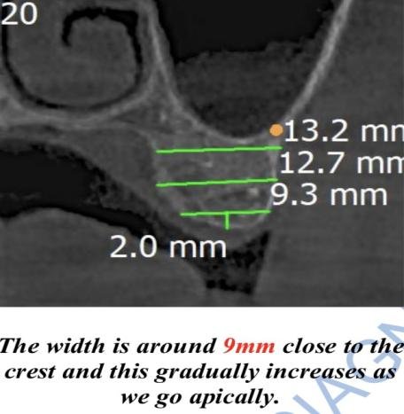
Fig 2
Preparation of implant site i.r.t 26
D3 bone density. Pilot drill bur was used in the clockwise direction with copious saline irrigation (Figure 3). Osteotomy site is supposed to be prepared 1mm short of the sinus floor, hence osteotomy site was prepared upto 6mm height. Osteotomy site was widened in the anti-clockwise direction with copious saline irrigation using the burs of the sizes 2mm, 2.3mm, & 2.5mm consecutively. Sinus floor was breached with the 3mm bur and Haptic feedback (jumping motion of the bur when it reaches the sinus floor) was felt (Figure 4 & 5). Next 3.3mm & 3.5mm burs were used to enter upto 3mm past the sinus floor (Figure 6). Novobone graft putty was placed using the gun. Do not push the graft but place gently (Figure 7, 8 & 9). The saline flow was stopped, 150rpm was set, 3.5mm bur was used to push the graft in the anti-clockwise motion (Figure 10 & 11). For molar 0.5cc of graft was placed. Radiovisiography (RVG) was recorded after graft placement. We can see radio-opaque ballooning in the sinus floor which indicates the graft placement in the elevated sinus floor (Figure 12). A Bioline implant of 8mm height & 4.2mm diameter was placed and RVG was recorded (Figure 13).
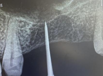
Fig 3
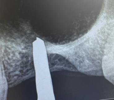
Fig 4
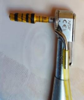
Fig 5
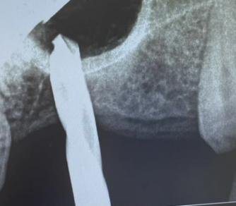
Fig 6

Fig 7
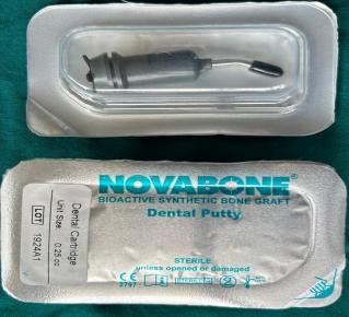
Fig 8

Fig 9
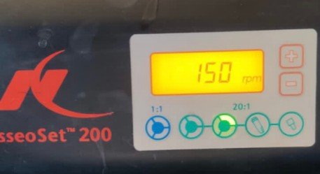
Fig 10
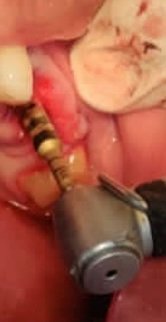
Fig 11

Fig 12
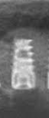
Fig 13
CBCT Images i.r.t 27 - Bone height: 7.5mm & width: 8mm (Figure 14 & 15)
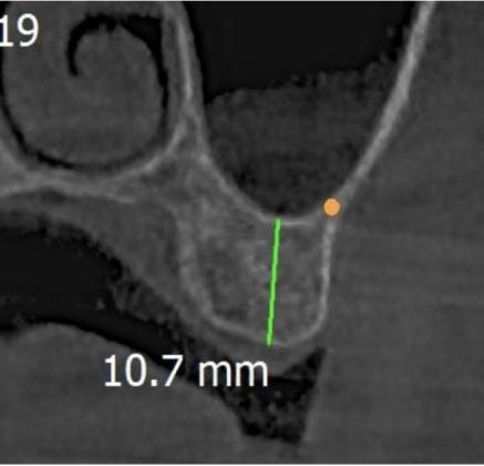
Fig 14

Fig 15
Preparation of implant site i.r.t 27
D3 bone quality. Pilot drill bur was used in the clockwise direction with copious saline irrigation and osteotomy site was prepared upto 6.5mm height (Figure 16). Osteotomy site was widened in the anti-clockwise direction with copious saline irrigation using the burs of the sizes 2mm, 2.3mm & 2.5mm consecutively. Sinus floor was breached with the 3mm bur and Haptic feedback was felt (Figure 17). Next 3.3mm & 3.5mm burs were used to enter upto 3mm past the sinus floor (Figure 18). 0.5cc Novobone graft putty was placed in a manner mentioned similar for 26 (Figure 19). Radiovisiography (RVG) was recorded after graft placement. We can see radio-opaque ballooning in the sinus floor (Figure 20). A Bioline implant of 8mm height & 4.2mm diameter was placed and RVG was recorded (Figure 21). Intra-oral site of implants placement (Figure 22).
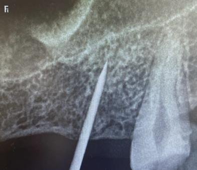
Fig 16
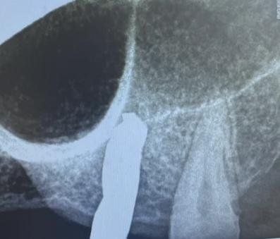
Fig 17
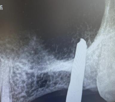
Fig 18
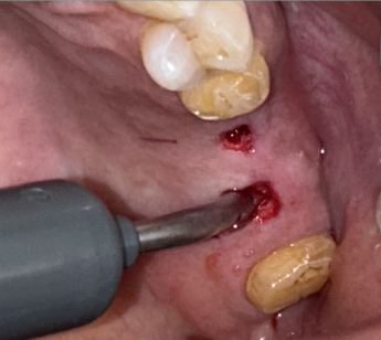
Fig 19

Fig 20

Fig 21
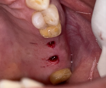
Fig 22
OPG recorded post implants placement
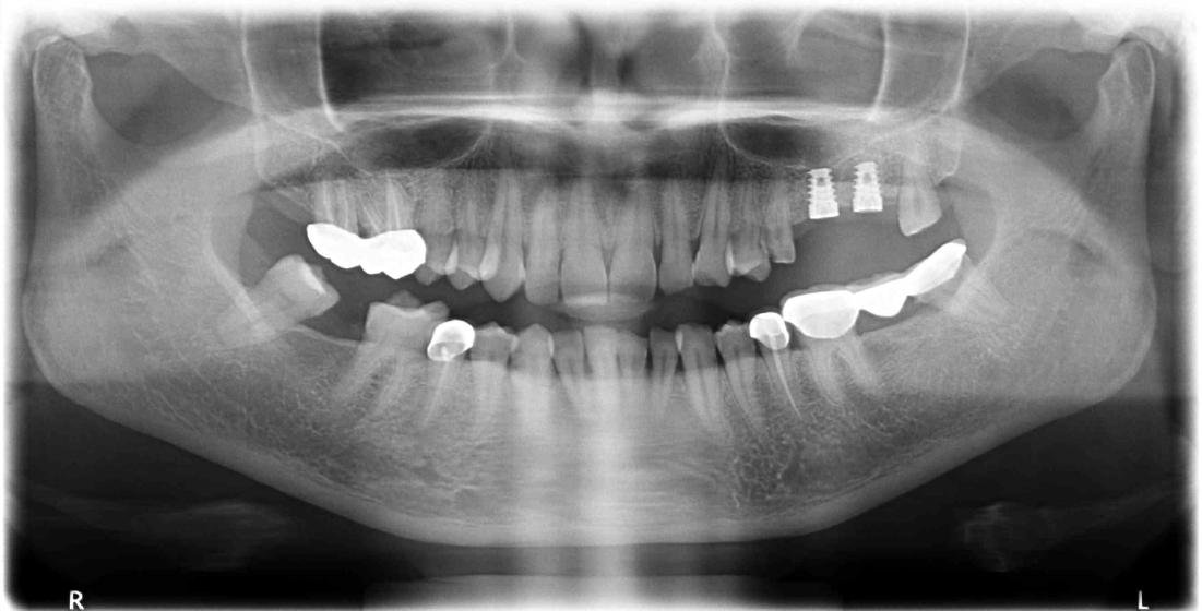
Fig 23

Fig 24: RVG-3 Months
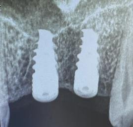
Fig 25: RVG-8 Months

Fig 26: Healing caps placed after 8 Months
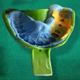
Fig 27: Light body & Putty impression

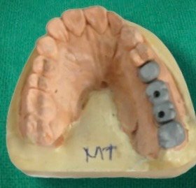
Jig trial
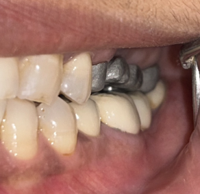
Metal trial

Final prosthesis
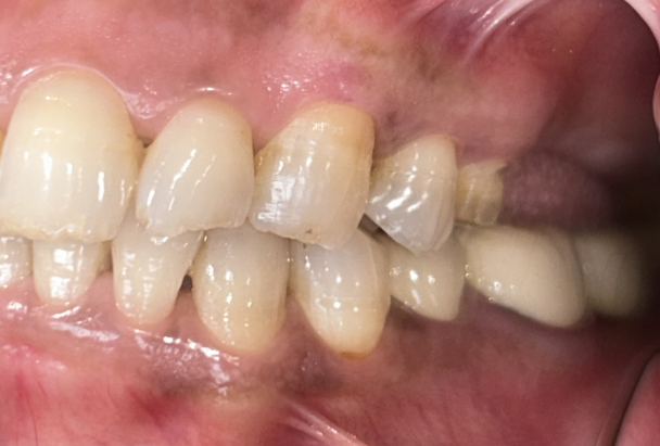
Pre Treatment
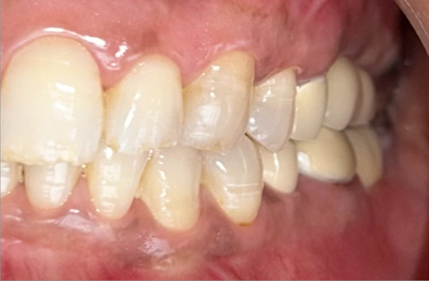
Post Treatment

Pre Treatment
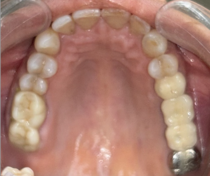
Post Treatment
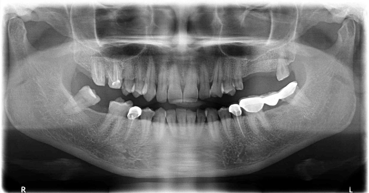
Pre Treatment OPG
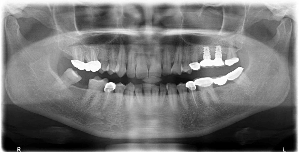
Post Treatment OPG
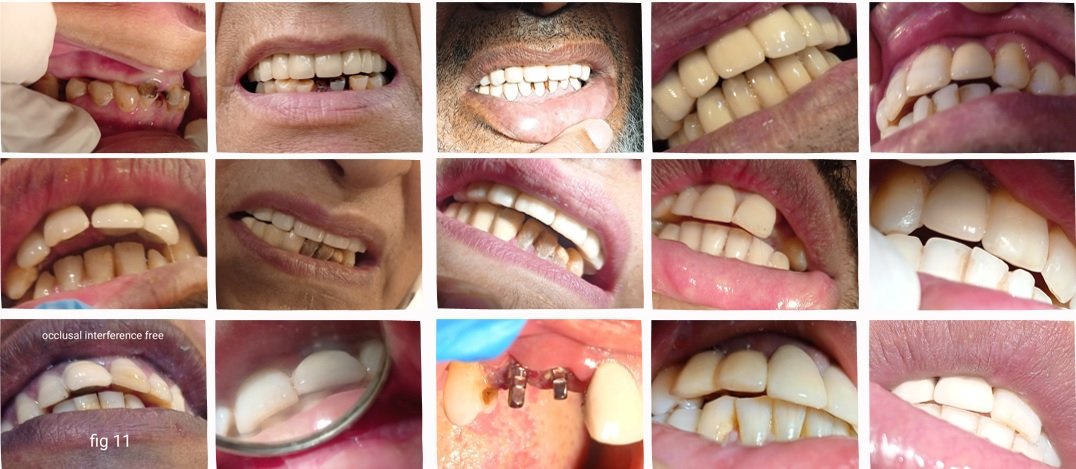
A metal ceramic crown was fabricated i.r.t 25 and a full metal crown i.r.t 28 due to decreased tooth height.
DISCUSSION
Trans-crestal sinus floor augmentation is a minimally invasive approach for placing implants in the posterior maxilla with reduced bone height with a low potential for complications. The use of Densah burs with their unique flute design and counterclockwise rotation allow for autogenous bone compaction laterally along the walls of the osteotomy as well as apically toward the sinus floor. This compaction together with modulated pressure during drilling and the constant in-and-out pumping motion ensures that this well hydrated autogenous bone creates a hydraulic pressure to gently lift and free the Schneiderian membrane. Further addition of a hydrated allograft material along with the propelling action of the burs gently pushes the graft against the membrane facilitating its lift. We believe that these unique features in the bur design and the concept of osseodensification have simplified the approach to a common problem in crestal sinus foor elevation.4 The quality and quantity of bone at the implant interface is linked to the success rate of osseointegration. The mechanical properties of bone are related not only to mineral density but also to the architectural distribution and collagen integrity. Collagen gives bone its toughness and its ability to dissipate energy. Therefore, collagen integrity has been found to be directly linked to bone plasticity. The plastic deformation of bone occurs as a gradual change that is dependent on time and strain rate. The fluid content of bone also plays an important role in determining bone viscoelasticity. Osseodensification is essentially a burnishing process that redistributes material on a surface through plastic deformation. The bur’s counterclockwise rotation causes the lands to slide across the surface of the bone with a compressive force less than the ultimate strength of the bone.5
CONCLUSION
Osseodensification is a novel, biomechanical, non-excavation osteotomy preparation method. Unlike traditional drilling, osseodensification utilizes proprietary high-speed densifying burs to compact and autograft bone in its plastic deformation phase. The result is an expanded osteotomy with preserved and condensed bone tissue that maintains alveolar ridge integrity and allows for implant placement with enhanced stability.6 Patients demand for a shorter duration and a faster final treatment. With the introduction of specially designed burs, making osseodensification possible, not only reduces treatment time but also gives a successful implant outcome. It is a promising concept which creates an autograft layer of condensed bone at the periphery of the implant bed with the use of densah burs that rotate in a clockwise and anti-clockwise direction, thereby enhancing implant stability and success. It is ideal for patients with poor bone quality, providing good primary implant stability.7
REFERENCES
- Samir J, Vijaysinh M, Rakhi P, Sudhir P, Krishnanunni N, Prashant P. Case Report Maxillary Ridge Augmentation Using Autogenous Block Bone Grafts Harvested From Symphyseal Region. Journal of Prosthodontics Dentistry. Jan to June 2020-Vol. 14, Issue 2, (Addendum-2), 70-78.
- Alhayati JZ and Al-Anee AM. Evaluation of crestal sinus floor elevations using versah burs with simultaneous implant placement, at residual bone height ≥ 2.0 _ < 6.0 mm. A prospective clinical study. Oral Maxillofac Surg. 2023 Jun;27(2):325-332.
- Karemore V, Phadnaik M, Dashputra A, Chodankar V. Crestal approach maxillary sinus augmentation by osseodensification with simultaneous implant placement. IP Int J Periodontol Implantol 2023;8(1):52-55.
- Shalash M, Mounir M, Elbanna T. Evaluation of crestal sinus floor elevation in cases exhibiting an oblique sinus floor with a residual bone height of 4.0-7.0 mm using Densah burs with simultaneous implant placement: a prospective clinical study. Int J Implant Dent. 2023 Nov 2;9(1):41.
- Huwais S, Meyer EG. A Novel Osseous Densification Approach in Implant Osteotomy Preparation to Increase Biomechanical Primary Stability, Bone Mineral Density, and Bone-to-Implant Contact. Int J Oral Maxillofac Implants. 2017 Jan/Feb;32(1):27-36.
- Huwais S. Enhancing implant stability with osseodensification-a case report with 2-year follow-up. Implantpractiseus.com
- Kanathila, Hema & Pangi, Ashwin. An Insight into the Concept of Osseodensification-Enhancing the Implant Stability and Success. Journal of Clinical and Diagnostic Research.7860/JCDR/2018/35626.11749.
Disclaimer from Guident
The views and opinions expressed in articles published in Guident are solely those of the authors and do not necessarily reflect the views of the editors, the editorial board or the publisher, Ivory India. Guident does not endorse any product, technique or treatment described. Clinical content is intended for qualified dental and medical professionals and is not a substitute for individual clinical judgement. Guident, its editors and the publisher accept no liability for any consequence arising from the use of information published in the journal.
How to cite this article
Dr. Brizita E. Kasturi, Dr. Subash M. Reddy, Dr. Sudhakar A.. Trans-Crestal Maxillary Sinus Floor Augmentation in Reduced Bone Height by Osseodensification, Followed by Bone Graft and Implants Placement: A Case Report. Guident. 2024 Sep;17(10). Available from: https://guident.in/articles/implantology/trans-crestal-maxillary-sinus-floor-augmentation-in-reduced-bone-height-by-osseodensification-followed-by.html