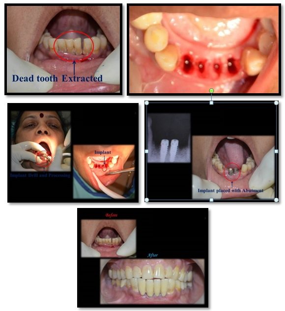
Implant Treatment in Esthetic Zone
Department of Prosthodontics, Teerthankar Mahaveer Dental College & Research Centre Moradabad, Uttar Pradesh, Department of Prosthodontics, New Delhi
ABSTRACT
Placement of implants in anterior aesthetic zone and to restore them remains a difficult area of implant dentistry. This may be attributed to the bone resorption that usually occurs after extraction of anterior tooth which, in turn alters the levels gingival tissue for the implant restoration. There has been a shift in focus from osseointegration, to creation of an implant borne restoration which is in harmony with the surrounding hard and soft tissue in the last few years. Thus the main objective remains the complete reconstruction of tooth and gingival-related aesthetics which, at times can be very difficult to attain.
Keywords: Dental Implants, Anterior maxilla, Customization Impression Copings.
INTRODUCTION
Achieving and maintaining optimal gingival esthetics around anterior implants is a demanding task. Inspite of the high success rates achieved with osseointegrated implants, high rate of gingival recession has been reported in anterior implant cases.1 The predictability of aesthetics depends on the loss of tissue present at the initiation of treatment. The greater the bone and soft tissue loss, the more difficult it is to produce an ideal aesthetic result. Esthetic success depends on a number of factors ranging from relationship between hard and soft tissues, soft tissue biotype and root position in the alveolar bone to three-dimensional implant position and angulation, and the contour of the abutment and the provisional restoration.2
CASE REPORT
A male patient aged 18 years, reported with missing maxillary central incisors to the private practice for the replacement of his missing teeth [Figure 1]. Patient gave a history of trauma and loss of teeth 8 months back. A thorough clinical examination was done followed by radiographic evaluations by taking intraoral periapical (IOPA) radiographs and orthopantomographs (OPG). Maxillary and mandibular diagnostic impressions were made to obtain diagnostic casts. A thorough medical history was taken to rule out any systemic disease. All the available prosthetic treatment options were explained. Bone mapping was done for the implant site and it revealed the labiopalatal cortical bone width to be 5.9 mm and mesiodistal length as 6.8 mm. It was planned to place an implant of size 4.0 mm × 11 mm in the left and right central incisor region. The patient was prepared for the surgery and local infiltration was given at site where implant was to be placed. Incision was given to elevate the mucoperiosteal flap. The osteotomy was started with a pilot drill initially and gradually, enlarged in width till 4.0mm and till a depth of 11 mm. During the osteotomy preparation paralleling pins were used to evaluate the parallelism of the implant site being prepared [Figure 2]. Once the osteotomy site was prepared, implant fixtures of dimension 4.0 mm × 11 mm were placed (Dentium NR Line,Korea ) and checked for parallelism radiographically [Figure 3,4]. Cover screws was placed and flap was sutured back with 4.0 non-resorbable silk sutures [Figure 5,6]. Postoperative instructions were given along with prescription of analgesics and antibiotics. Follow-up was done after every 2 weeks. After 4 months, the second stage surgery was done in the central incisor region and a gingival formers of size 4.5 mm × 3 mm were placed. After 1 week the suture placed were removed and gingival formers were replaced by closed tray impression posts which were customized with flowable composite according to soft tissue architecture[Figure 7]. A customized closed tray impression was made with elastomeric impression material. Zirconia crowns were fabricated and cemented. Chair-side postoperative IOPA radiograph was taken to assess the complete seating of implant abutment on implant fixture. The patient’s occlusion was checked and the implant retained fixed prosthesis was evaluated for esthetics and phonetics [Figure 8].

Figure 1: Pre-Operative

Figure 2: Paralleling Pins in Place
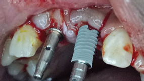
Figure 3: Implant Fixture Placement (Dentium NR Line,Korea )
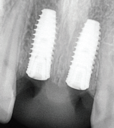
Figure 4: Post Implant Placement IOPA

Figure 5: Cover Screw Placement
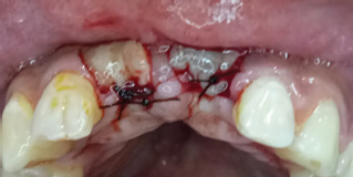
Figure 6: Sutures placed
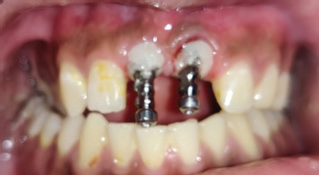
Figure 7: Customised Impression Copings

Figure 8: Final Prosthesis
DISCUSSION
Replacement of missing maxillary anterior teeth with dental implants is a viable treatment option. Dental implant-supported prosthesis highly beneficial over crown and bridge or removable prosthesis. Dental implants help in retaining residual bone, ease of oral hygiene maintenance, and non-involvement of adjacent teeth.3 Certain clinical parameters play an important role in order to provide successful and aesthetic treatment. This is specifically true in the anterior maxilla, where the teeth and their supporting structures are readily visible.4 Successful implant treatment to replace missing teeth in the anterior maxilla requires good preoperative planning and a precise surgical plan, and ultimately prostheses are fabricated in consideration of function and soft-tissue support.4,5,6 Pre planning of the prosthesis is an important criteria to achieve desired end results. Since the use of fixed partial dentures necessitates the destruction of adjacent teeth to prepare them as abutments and loss of valuable tooth structure it is not a great choice especially in young patients. Removable partial dentures leads to the loss of alveolar bone on both abutment and non-abutment teeth. Moreover the dissatisfaction rate of removable partial dentures is also relatively high.
CONCLUSION
It becomes imperative to do proper planning, precisely place the implants and handle the soft tissue delicately if the anterior teeth need to be restored successfully with implants. Timing of placement, subcrestal positioning, and choice of implant system are also important to get it right in the esthetic zone.
REFERENCES
- Gotfredsen K. A 10-year prospective study of single tooth implants placed in the anterior maxilla. Clin Implant Dent Relat Res. 2012;14(1):80–7.
- Kan JY, Rungcharassaeng K. Site development for anterior single implant esthetics: The dentulous site. Compend Contin Educ Dent. 2001;22(3): 221–6. 228, 230-1.
- Touati B, Guez G, Saadoun A. Aesthetic soft tissue integration and optimized emergence profile: provisionalization and customized impression coping. Pract Periodontics Aesthet Dent. 1999;11: 305-316.
- Phillips K, Kois JC. Aaesthetic peri-implant site development. The restorative connection. Dent Clin North Am 1998; 42: 57–70.
- Sonick M. A comparison of the accuracy of periapical, panoramic and computed tomographic radiographs in locating the mandibular canal. Int J Oral Maxillofac Impl. 1994;9: 455–60.
- Kois JC. Predictable single tooth peri-implant aesthetics: Five diagnostic keys. Compendium 2004; 25: 895–905.
Disclaimer from Guident
The views and opinions expressed in articles published in Guident are solely those of the authors and do not necessarily reflect the views of the editors, the editorial board or the publisher, Ivory India. Guident does not endorse any product, technique or treatment described. Clinical content is intended for qualified dental and medical professionals and is not a substitute for individual clinical judgement. Guident, its editors and the publisher accept no liability for any consequence arising from the use of information published in the journal.
How to cite this article
Dr. Abhishek Sharma, Dr. Deepanshi Garg. Implant Treatment in Esthetic Zone. Guident. 2020 Feb;13(3). Available from: https://guident.in/articles/implantology/implant-treatment-in-esthetic-zone.html