Guided Implant Surgery
A.B Shetty Memorial Institute of Dental Science, Nitte University, Deralakatte, Mangalore, India
Abstract
Implant treatment is not only the first choice for a dental professional but also highly sought by the patients themselves due to heightened awareness among the general population. Optimal placement of dental implant is the basic requirement for the success of implant treatment. In the present day of technology, guided implant surgery is only logical to use the best imaging technique for placing dental implants to perfection. Rather than following the traditional surgical procedure which puts us at disadvantage of not knowing the exact location and position of the planned implant, guided surgery makes our chances of successful implant placement much brighter by making the adjacent anatomic structures much easier to negotiate.
INTRODUCTION
The success of implant therapy mainly depends on appropriate treatment planning and properly performed implant placement surgery. The success of implant-supported restorations is not only depended to the implant integration level in the bone but also to the implant position. The conventional treatment of the patient with dental implants includes a diagnosis, treatment planning, a diagnostic waxing, and a surgical template to place implant. The position of implants placed depends on radiographic diagnosis, anatomical landmarks, the quality and quantity of bone, and biomechanics. But transferring the exact position of the planned implants to the patient can be difficult, imprecise, and require extensive laboratory procedures.123
Guided implant surgery uses patient’s CT images and 3-dimensional surgical planning software to plan implant surgery. Then planned position of implants is transferred to the patient with a surgical template. This procedure may increase success of immediate loading for the partially edentulous patient. Whereas, Conventional implant planning with panoramic x-ray images has its limitations because precise execution of planned surgery is very difficult.4-7
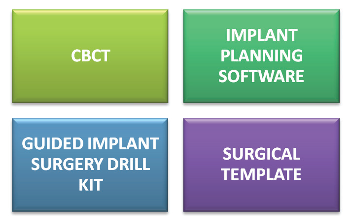
Guided implant surgery can be executed accurately and precisely as planned. For guided implant surgery; a customized surgical template and a specialized surgery tool kit is required.
TOOLS USED IN GUIDED IMPLANT SURGERY
SURGICAL TEMPLATE
Surgical template is equipment that holds the surgical planning data. It is worn by the patient during surgery. On the surgical template, small metal implant positioning sleeves are placed, which helps to guide the drilling and implant direction. It is necessary to fabricate surgical template to perfectly fit the patient.
The CAD/CAM surgical template can precisely transfer the planned positions from the software to the patient with the use of surgical instruments and, therefore, there is a increase chance of having a more stable implant. surgical guide can be of bone supported, mucosa supported, tooth supported.
GUIDED IMPLANT SURGERY KIT
Guided implant surgery kit includes anchor pin /anchor drill, tissue punch, drill guide. Anchor pins hold the surgical template firmly fixed to the oral cavity. Drilling is done with the Anchor Drill then Anchor Pin is inserted to place the surgical template. Tissue punch is used to remove tissue during flapless implant surgery. Drill guide attaches to the sleeve and it helps to guide the drill into the planned angle and direction. There are different drill guides to accept different drill sizes.
CBCT and implant planning software
Cone-beam CT is specialized x-ray equipment that scans the patient in three-dimension by using Cone-beam CT along with proper software. thus a precise and accurate examination of the patient, with realistic visuals, can be achieved Cone-beam CT produces images in DICOM format, and a 3D implant planning software reads these files and reconstructs them in two-dimensional or three-dimensional images. These software provide various tools to plan implant position, thus the user can refer to the anatomic structure of the patient and plan a safe surgery.8
PROCEDURE
Guided implant surgery is mainly subdivided in following step.

CT scanning
CT scanning with the correct parameters is the basis for accurate planning in the software and for correct implant placement. To get the optimal scan data, the radiologist and the patient need to be instructed correctly and scanning instructions or parameters must be followed according to the computer software.
Software-based planning and fabrication of the surgical template
Planning with the help of computer software allows for implants to be planned virtually within the planning software. When it completed, the case plan is sent to the surgical template manufacturer. Either the software company may act as the surgical template manufacturer or the dental laboratory may fabricate the surgical template according to the software concept used.
Surgery with guided template & guided implant insertion
Surgical template is equipment that holds the surgical planning information. On the surgical template, small metal sleeves are present, which guides the drilling and implant direction. It is necessary for the surgical template to be fabricated to precisely fit the patient. After placement of surgical template on the ridge; implant placement is carried out.8,9
DISCUSSION
CT/CBCT scanners make possible for the dentist to visualize a patient’s anatomy in 3 dimensions. With the CT/CBCT visualization of the exact height and width of available bone for implant placement, soft tissue thicknesses, the exact location of the maxillary sinuses, proximity and root anatomy of adjacent teeth and other important vital structures such as the mandibular canal, mental foramen, and incisive canal are possible10-12. these images can be used with proprietary software programs to virtually plan the placement of implants for an individual patient’s anatomy and case plan. Thus the size of the planned implant, its position within the bone, and its proximity to vital structures can be checked before surgery.12-14 Then computer-generated surgical drilling guides can be fabricated from the virtual treatment plan. And then dentist can use these surgical guide to place the planned implants in the patient’s mouth in the same positions as in the virtual treatment plan, resulting in a more accurate and predictable implant placement and decrease the chance of implant failure.15-17
There are three types of computer-generated surgical guides available: tooth supported, mucosa supported, and bone supported. Tooth-supported guides are generally used in partially edentulous patients. The surgical guide is designed in a way that it rest on other teeth in the arch for accuracy of guide fit. Mucosal-supported guides are used mainly in edentulous patients and it rest on the mucosa. Bone-supported surgical guides can be used in both partially or fully edentulous cases, but are used mainly in fully edentulous patients in which significant ridge atrophy is present and accurate fitting of a mucosa-supported guide is questionable. These guides include elevation of an extensive full-thickness flap to expose the bone in the planned implant sites as well as in the adjacent areas for stable seating of the guide over the bony ridge.18
The manufacture of a stereolithographic surgical guide or model involves reproducing the digitally planned dimensions of the surgical guide or model by using a laser beam to selectively solidify an ultraviolet-sensitive liquid resin. Stereolithographic materials have inherent potential problems that can lead to light sensitivity and expansion and/or shrinkage of the material. Leaving them exposed to light for extended periods of time, as well as sterilization in high-temperature autoclaves, distorts stereolithographic materials. The literature concludes that implant site preparation using surgical drill guides generates more heat than classic implant site preparation, regardless of the irrigation system used19
Dental implant placement using CT-guided surgery with drill guides is known to enhance safety compared with using a freehand technique. However, no available CT-guided drill guide technology exists today with absolute precision. All articles on stereolithographic guides show error in all dimensions between virtual planning and obtained implant positions. According to the literature, implants placed by bone-supported guides have the highest mean deviations, whereas implants placed by mucosa-supported guides have lower deviations.48 Tooth-supported guides have the lowest measured deviation. A single guide, using metal guide sleeves and rigid screw or pin fixation with specific drilling instrumentation, further minimizes error. Most systems use these fixation techniques to stabilize mucosal-supported guides.20-22
Compromised patients such as Pre- and postoperative radiation therapy potentially alter healing capacities in patients who have head and neck cancers. The literature advocates pre- and postoperative courses of hyperbaric oxygen therapy (HBO) to increase bone vascularity before implant placement in these patients.23-25 Placing implants with minimal flap elevation and hard and soft tissue trauma is indicated in these patients to minimize the likelihood of the development of osteoradionecrosis. Bleeding, swelling, and alteration of the blood supply can be reduced by using these technologies.26,27 Patient’s historical experiences in dental offices vary greatly. Procedures that require long periods in a dental chair can prevent some patients from seeking treatment because of extreme levels of anxiety, stress, and phobias. Orthopedic and spinal disorders may limit the amount of time a patient can sit in a dental chair. Wheel chair bound patients pose another set of logistical treatment problems. These types of patients can require extensive planning and preparation before treatment. Treatment must be performed quickly and efficiently, without compromising quality. Computer generated implant planning allows for the preoperative visualization of most of the potential planning and anatomic issues that may be encountered during surgery, all before the patient sits in the dental chair. By using surgical guides, implants can be placed quickly and predictably, thus minimizing patient stress, pain, and time in the dental chair.
Thus basic advantages of this procedure includes (A) shorter surgery times, (B) less invasive, flapless surgery and, thus, less swelling, less pain, and shorter initial healing times, (D) can use of the fixed prosthesis immediately.8 CT-based technologies available today have their own limitations and certain limitations of computer guided surgery include the additional cost of the planning software, special surgical inventory, and fabrication of the stereolithographic template, difficult to deal with unexpected situations during surgery.8 Chanseop Park et all did a study on measurement and comparison of the accuracy of posterior implant placement using 3 precision surgical guides with different occlusogingival heights, and to evaluate the difference in accuracy of implant placemen with precision guides as compared to freehand placement. And they concluded that surgical guides with 4-mm occluso gingival height allow placement as accurate as surgical guides with 8-mm height. Placement through the guide achieved the target position more accurately than freehand insertion. 28
Andreas Pettersson et all did a study on cadavers to check Accuracy of CAD/CAM-guided surgical template implant surgery. And they found that demonstrated a statistically significant difference between mandibles and maxillae for the hex, apex, and depth measurements in the variation between the virtually planned implant positions and the positions of the implants placed after surgery with a CAD/CAM-guided surgical template.29
Timo Kero et all did a study on cadavers to perform virtual variation simulations on virtually planned implant placements and to compare them with corresponding results from actual surgeries performed on human cadavers in a previous study.30
CONCULSION
With the advent in diagnostic imaging and acceptance of implants as a prosthodontics modality there has been an emergence of increased utilization of imaging techniques for favourable placement of implant. However several unexpected procedure linked adverse events during guided implant surgery indicate that the clinical demands on the surgeon were no less than those during conventional placement.
REFERENCES
- Adell R, Lekholm U, Rockler B, Branemark PI. A 15-year study of osseointegrated implants in the treatment of the edentulous jaw. Int J Oral Surg 1981;10:387-416.
- Rangert B, Jemt T, Jorneus L. Forces and moments on Branemark implants. Int J Oral Maxillofac Implants. 1989;4:241-7.
- van Steenberghe D, Ericsson I, Van Cleynenbreugel J, Schutyser F, Brajnovic I, Andersson M. High precision planning for oral implants based on 3-D CT scanning. A new surgical technique for immediate and delayed loading. Appl Osseointegration Res 2004;4:27-31
- van Steenberghe D, Naert I, Andersson M, Brajnovic I, Van Cleynenbreugel J, Suetens P. A custom template and definitive prosthesis allowing immediate implant loading in the maxilla: a clinical report. Int J Oral Maxillofac Implants 2002;17:663-70.
- van Steenberghe D, Glauser R, Blomback U, Andersson M, Schutyser F, Petersson A, et al. A computed tomographic scan-derived customized surgical template and fixed prosthesis for flapless surgery and immediate loading of implants in fully edentulous maxillae: a prospective multicenter study. Clin Implant Dent Relat Res 2005;7 Suppl 1:S111-20
- Marchack CB, Moy PK, the use of custom template for immediate loading with the definitive prosthesis: a clinical report. J Calif Dent Assoc 2003;31:925-29.
- .Marchack CB. An immediately loaded CAD/CAM-guided definitive prosthesis: a clinical report. J Prosthet Dent 2005;93:8-12
- Introduction of guided implant surgery; CYBERMED INC., ONDEMAND3D TECHNOLOGY INC.
- Christopher B. Marchack, CAD/CAM-guided implant surgery and fabrication of an immediately loaded prosthesis for a partially edentulous patient; J Prosthet Dent 2007; 97: 389-394
- Ramez J, Donazzan M, Chanavaz M, et al. The contribution of scanner imagery in implant surgery and sinus overflow using frontal oblique orthogonal reconstruction. Rev Stomatol Chir Maxillofac 1992;93:212–4.
More references are available on request
Disclaimer from Guident
The views and opinions expressed in articles published in Guident are solely those of the authors and do not necessarily reflect the views of the editors, the editorial board or the publisher, Ivory India. Guident does not endorse any product, technique or treatment described. Clinical content is intended for qualified dental and medical professionals and is not a substitute for individual clinical judgement. Guident, its editors and the publisher accept no liability for any consequence arising from the use of information published in the journal.
How to cite this article
Dr. Manoj Shetty, Dr. Krishna Prasad D, Dr. Aadit B. Patel Department of Prosthodontics. Guided Implant Surgery. Guident. 2015 Feb;8(3). Available from: https://guident.in/articles/implantology/guided-implant-surgery.html