Diagnostic and Imaging Techniques in Implant Dentistry
Department of Prosthodontics, Crown, Bridge and Implantology, AME’s Dental college and Hospital, Raichur
ABSTRACT
The diagnostic imaging in implant dentistry has commuted drastically over the years. Traditionally, the use of two- dimensional imaging techniques were used. But with the increased demand of implants, its accurate positioning, in the exact site has led to development of various changes in the type of imaging techniques used in each scenario leading to greater implant success.
This article reviews the various Diagnostic imaging techniques its associated advantages ,disadvantages and the precise timing that each imaging technique should be used will be discussed.
Key Words: Imaging, implant, techniques
INTRODUCTION
The decision to image the patient is based on the patient’s clinical needs. To obtain a radiologic survey of a particular area of interest, the imaging modality is selected that yields the necessary diagnostic information related to the patient’s surgical and prosthetic needs. Maximizing the ratio of benefit to risk for acquiring information from imaging examinations is a fundamental tenet of radiology. Examinations known to produce this result are not necessarily the examinations that cost the least, are in proximity to the dentist, or produce the lowest radiation exposure.1 However, they enable the implant team to acquire necessary information that is crucial to the success of the implant procedures.1
CLASSIFICATION OF IMAGING MODALITIES BASED ON PHASES OF USE 1,2
- PHASE 1 -Pre prosthetic Implant Imaging
- PHASE 2 -Surgical and Interventional Implant Imaging
- PHASE 3 – Post Prosthetic Implant Imaging
The objective of this phase is to identify any disease, determination of bone quality, quantity, implant position and orientation.
evaluate the healing and integration of surgery sites. The correct position of the abutment and prosthesis fabrication are ensured in this phase.
This phase starts from the placement of the implant and lasts as long as the implant remains in the jaw. The radiographic sequence for phase III imaging is post-prosthetic imaging, followed by recall and maintenance imaging and an evaluation of alveolar bone change.
IMAGING MODALITIES can be subdivided into the following 1
- Planar Two dimensional
- Quasi three dimensional
- Three dimensional
Includes
1.1) Periapical
1.2) Bitewing
1.3) Occlusal
1.4) Cephalometric
2.1) Tomography
2.2) Panoramic imaging
3.1) CT
3.2) MRI
1.1) PERIAPICAL RADIOGRAPHY1,3: Periapical radiographs are images of a limited region of the mandibular and maxillary alveolus.
Principle: produced by placing the film intraorally parallel to the body of the alveolos with the central ray of the X ray device perpendicular to the alveolus at the region of interest, producing a lateral view of the alveolus.
ADVANTAGES
- Readily available to most dentists
- Delivers fairly low effective dose of radiation (below 5 microseiverts)3
- Useful high yield modality for ruiling out local diseases.1
- Identifies critical structures1
DISADVANTAGES 1
- Limited value in determining quantity, because the image is magnified and distorted.
- Does not depict 3 dimension of bone width
- Limited value in determining bone density (because lateral cortical plate prevent accurate interpretation)
- Provides lateral view but no cross sectional information.
USES 1
- In Pre prosthetic phase: single tooth implant region with abundant tooth width.1
- In surgical phase: immediate feedback after osteotomy preparation especially if next to existing natural tooth or any vital structures.3
- In post prosthetic phase: for evaluating changes in bone mineralization or bone volume adjacent to the placed implants.

Periapical radiograph at implant placement

Periapical radiograph at the definitive prosthesis placement
1.2 BITE WING RADIOGRAPH 4
The bitewing radiograph is an image that depicts the maxillary and mandibular crowns of the teeth, providing clear image of the interproximal surfaces of the teeth.
ADVANTAGES 4
- Useful in identifying significant periodontal bone loss around implants
- Interproximal dental decay especially in adjacent teeth

Vertical bitewing radiograph of the implant-supported crown. Slight crestal bone loss evident. Radiographic appearance of commonly used cements in dentistry
1.3 OCCLUSAL RADIOGRAPH 1,5
Intraoral occlusal radiographs are capable of demonstrating almost the entire alveolar process of either the maxilla or the mandible.
High-resolution planar images of the mandible or the maxilla are produced by occlusal radiography.
ADVANTAGES
- Structures like maxillary sinus, nasal cavity, and nasopalatine canal can be assessed through occlusal radiography
- As periapical radiographs are unable to produce any cross-sectional information, occlusal radiographs are sometimes used to determine the facio-lingual dimensions of the mandibular alveolar ridge.
- Helps in evaluating extents of lesion, cysts and malignancies.10
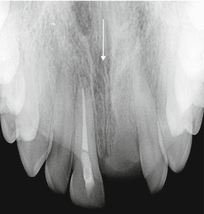
Nasopalatine canal seen from maxillary occlusal view

Periapical radiograph at implant placement
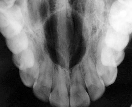
Periapical radiograph at the definitive prosthesis placement
DISADVANTAGE
- a true cross-sectional view in the axial plane, unobstructed by superimposition, is possible only in the mandibular arch.
- the mandibular occlusal radiograph at best describes the maximum width of bone, which is usually at the base of the jaw, and fails to show the medial and lateral extent of cortical bone delineating the alveolar process and the salivary gland fossa.6
- Causes superimposition of images2
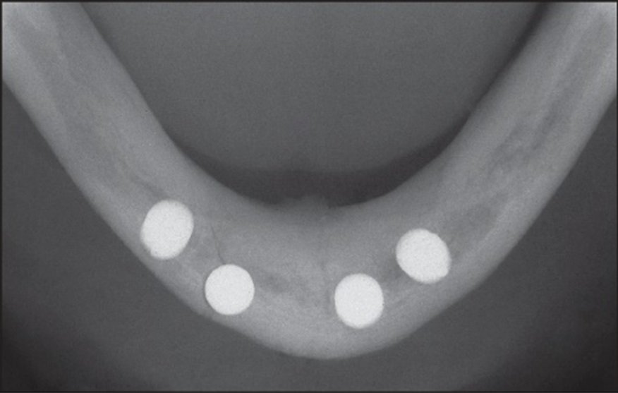
Post operative occlusal radiograph showing the implants in the mandibular anterior region7
1.4 CEPHALOMETRIC RADIOGRAPHS
Although low cost and availability are advantages, when this technique is used for implant planning, the advantages are limited.
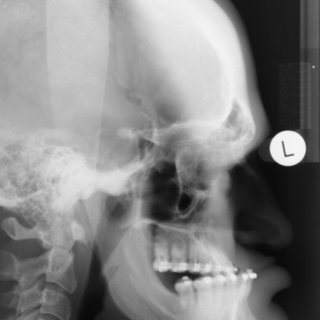
Either lateral or A-P evaluation can be useful in treatment planning for implants.8
ADVANTAGES
- They provide angulation, bone height and thickness and soft tissue profiles.
- They offer relationship of maxilla to mandible
- Orthognathic surgery may be recommended before implant reconstruction to relieve the increased occlusal forces often present when restoring patients without a class I jaw relation .
- they outline the geometry of the alveolus in the anterior region and the relationship of the lingual plate to the patient’s skeletal anatomy1

Image appreciating soft tissue profile and maxillomandibular relations

DISADVANTAGES 9
- The visualization of most significant details is limited in cephalometric radiography by the overlapping of the left and right sides
- Because of the overlapping bone structures, measurements of bone height and width in the symphyseal area may mask local bone defects
- Overly optimistic bone volume assessments are created due to the presence of genial tubercles
- Images do not provide useful information when planning placement of implants lateral to the mid-sagittal plane
- not useful for demonstrating the bone quality and only demonstrates a cross-sectional image of the alveolus where the central rays of the X-ray device are tangent to the alveolus.2
- First, only the anterior mandible is visible in cross-sectioning. In both the anterior and lateral views, a majority of the maxilla and mandibular dentition and osseous structures overlap, producing distortion and inaccuracy of measurement. Patient positioning is also difficult to reproduce for purposes of comparison.


QUASI THREE DIMENSIONAL IMAGING MODALITIES
1. PANORAMIC IMAGING
Panoramic radiography gives a general overview of the jaw bones and relevant anatomic landmarks. For more detailed bone images, intraoral radiographs are normally taken to complement the examination.9
ADVANTAGES
- Opposing landmarks are easily identified.
- Vertical height of bone initially can be assessed
- Convenience of procedure
- Gross anatomy of jaws and any related pathology findings can be evaluated.
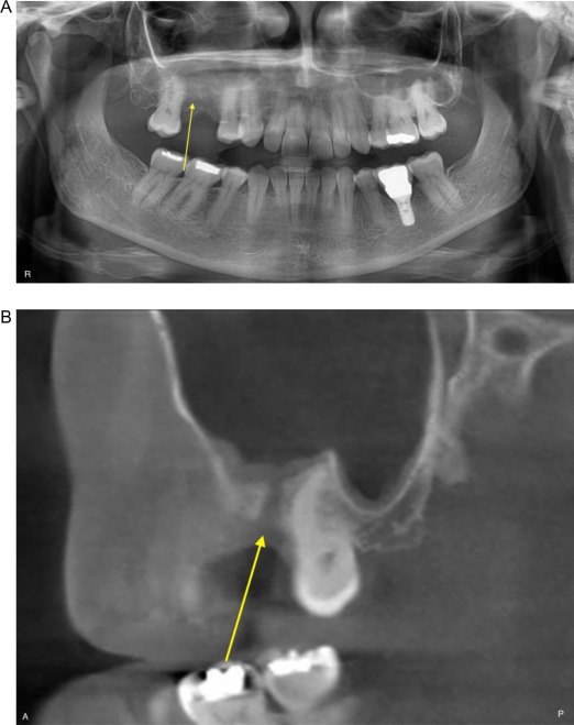
Maxillary sinus showing close proximity to the planned implant site thereby helping in initial assessment of implant region
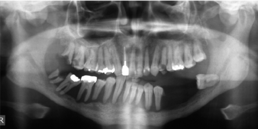
Panoramic radiography showing resorption and decreased height of bone for placement of implants

Pre-operative panoramic radiograph of residual cyst in the mandible with notable thinning of the mandibular cortex
DISADVANTAGES
- Does not demonstrate bone quality/mineralization
- Misleading quantitatively because of magnification and because third dimension cross sectional view is not demonstrated.
- TOMOGRAPHY: In Greek Terms Tomo means slice and graphy means picture .Tomography was first adopted in the year 1962 by International Commission on Radiological units and measurements of all forms of body section radiography.
BASIC PRINCIPLE
It selects a level in the body and blurs out structures above and below that plane leaving a clear image of this selected image .
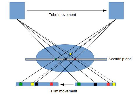

VARIOUS MOVEMENTS IN TOMOGRAPHY
- LINEAR
- CIRCULAR
- FIGURE 8
- HYPOCYCLOIDAL
- ELLIPTICAL
- SPIRAL
Linear movements is a two dimensional whereas the rest of the motions are called complex tomographic movements
ADVANTAGES OF TOMOGRAPHY 1,2
- Taking magnification into consideration it enables quantification of geometry of alveolus
- enables determination of spatial relationship between critical structure and implant site.
DISADVANTAGES
- Not particularly useful in determining bone quality or identifying dental and bone disease.
THREE DIMENSIONAL IMAGING MODALITIES
- Computed tomography
Computed Tomography was invented by Sir Hounsefield and was announced to the imaging world in 1972. CT is a digital and mathematical imaging technique that creates tomographic sections where the tomographic layer is not contaminated by blurred structures from adjacent anatomy. Here the radiologist could view hard and soft tissues on an image without performing an invasive procedure .
Principle:1 CT produces axial images of patients anatomy. Axial images are produced perpendicular to the long axis of the body.
X ray source is attached rigidly to a fan beam geometry detector array which rotates 360 degree around patient. And collects data.
Speciality of CT Images 1
CT images are inherently 3D images typically 512*512 pixels with a thickness described by the slice spacing of imaging technique .The individual element of CT image is called voxel, which has a value referred to in Hounsfield unit.
Advantages: 1,2,9
- Enables reformatting the image data to create tangential and cross-sectional tomographic images of implant site.
- The density of structure is absolute and quantitative and can be used to differentiate tissues in the regions and characterize bone quality.
- Identification of critical structures at the proposed region.
- Enables determination of position and orientation of dental implants.
- CT preserves soft tissue details which might be useful when reconstruction is to be done in an aesthetic area.
- Bone density can be measured on CT data and this is important in assessing bone quality especially upper jaw which has in general lower bone density.
- Using CT ,the jaws can be seen virtually from any pt on screen and implant placement can be done interactively.
- Provides 3- dimensional multiplanar view ,accurate dimension
- Optimal resolution of potential implant site and surrounding structures .
Disadvantages
- Total radiation dose of CT is much higher than compared to conventional CT scan
- The costs of CT is higher
- Limited availability of CT scanners
- Technical errors
- Need for special training in image interpretation.
CONE BEAM COMPUTED TOMOGRAPHY 11
CBCT is an advanced digital imaging technique that allows the operator to generate multiplanar slices of a region of interest and to reconstruct a 3D image of these structures of interest by using a cone-shaped rotating x-ray beam via a series of mathematical algorithms.

ADVANTAGES OF CBCT 11
- Mutiplanar reconstruction
- Significantly less radiation compared with other 3D advanced imaging modalities (ie, medical CT)
- Fast, efficient, in-office modality
- Interactive treatment planning
- Adequate for bone grafting assessment
- Computer-aided surgery

Different possible views of TMJ & Maxillomandibular relation by using CBCT
DISADVANTAGES OF CBCT
- Limited soft tissue visualization
- Some CBCT machines produce an increased radiation exposure compared with selected intraoral and panoramic radiographs
- Limited bone density measurements
- Artifacts created by metal subjects (eg, PFM crowns, dental implants), costly
- Third-party software applications and 3D models are an additional expense
The adjunctive use of CBCT in implant dentistry can be divided into four main categories:11
- Diagnostics
- Implant planning
- Surgical guidance
- Postimplant and/or post grafting evaluation
CBCT AND DIAGNOSTICS
- evaluation of the proposed implant site to exclude the presence of occult pathology, foreign bodies
- to determine the suitability of the site in terms of 3D morphology and proximity to vital anatomical structures.
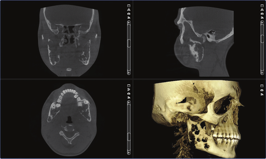
CBCT imaging of a pathologic lesion located in the mandible (reparative giant cell granuloma)15
CBCT AND IMPLANT PLANNING
- one of the most frequently reported applications of CBCT is linear measurement of the ridge. Magnification of linear measurements does not occur in CBCT.
- CBCT images provides reliable bone quantity information for preoperative implant planning in different areas of the maxilla and mandible.
- ability to evaluate the ridge topography and proximity to vital anatomical structures three dimensionally for grafting purpose.
- CBCT can accurately assess the thickness of cortical bone such as the facial/buccal and lingual/palatal cortical plates, the floor of the nasal cavity, and the medial and lateral walls of the maxillary sinuses.
- Evaluation of bone density has also been an area of increasing interest. To date, it has been possible to obtain only relative bone quality information. However, several research studies have been done to assess the reliability of bone density measurements obtained with CBCT in an effort to overcome this limitation. The use of the quantitative CBCT (QCBCT) method holds promise as an alternative diagnostic tool for preoperative bone density evaluation.
- Effective in locating blood vessels in the lateral wall of the maxillary sinus and mandibular symphysis region which should be appreciated before sinus augmentation procedures. And for avoiding perforations respectively.
CBCT AND SURGICAL GUIDANCE
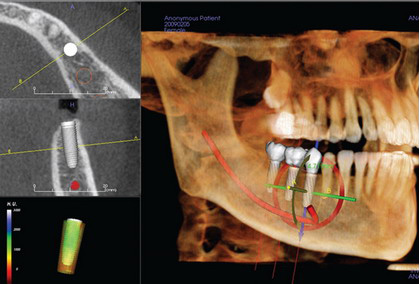
- As treatment planning software where, virtual implants are simulated as a precursor to the fabrication of surgical guides that will be used at the time of implant placement.
- Active CBCT-aided implant surgery refers to the use CBCT data and surgical navigation systems to perform fully computer-guided implant placement. The accuracy of navigation systems has been tested in some studies; however, more research is need in this area.
MAGNETIC RESONANCE IMAGING 1,12
MRI is a technique developed in medical in medical imaging that is probably the most innovative and revolutionary other than CT. It is a technique to image the protons of the body using magnetic fields , radiofrequencies, electromagnetic detectors and computers.
MRI Images :-Characterized by voxels with an in-plane resolution measured in pixels (512*512) and millimetres and a section thickness measured in millimetres (2to 3mm) for high resolution imaging acquisitions .Resulting MRI images are the antithesis of CT images with cortical bone appearing dark or black and fat or water appearing bright or white.

USAGE OF MRI FOR DENTAL IMPLANTS
MRI is used in implant imaging as a secondary imaging technique when primary imaging techniques such as complex tomography, CT or ICT fail.
Complex tomography fails to differentiate the inferior alveolar canal in about 60% of cases and and CT in about 2% and this could be attributed to the osteoporotic trabecular bone and poorly corticated inferior alveolar nerve .MRI visualizes the fat in trabecular bone and neurovascular bundle in adjacent trabecular bone.
ADVANTAGES OF MAGNETIC RESONANCE IMAGING 12
- Gives suitable information on the 3 D relationships of all relevant anatomic structures.
- In a study conducted by Gray CF et al. it was seen that MRI is reasonably tolerant of artefacts caused by metal pins and amalgam fillings.
- Unlike CT and other X Ray tech, MRI uses no ionizing radiation.
- MRI gives good bone detail because cancellous bone yields a strong signal from the marrow fat.
- Excellent anatomic detail provided by thin slice high resolution MRI allows for assessment of suitability of sites to receive an implant in terms of bone quality and thickness and important structures such as Inferior alveolar nerve and sinus.
DISADVANTAGES OF MRI IN DENTAL IMPLANT IMAGING
- MRI is not useful in characterizing bone mineralization or as a high yielding technique for identifying bone or dental disease.
- Detection of hard tissues (eg, enamel, highly mineralized cortical bone) is compromised with MRI because of low concentration of free H+ ion in solution.
- MRI cannot be scrutinized as predominant diagnostic tool used for imaging.
A study conducted by Laura et al13 compared CBCT and MRI for the superiority in diagnosing nerve lesion associated with implant placement and it was noted that MRI enabled a precise depiction of implant position and its spatial relation to the IAN ,and thus offering an objective assessment of nerve injuries by direct visualization of inferior alveolar neurovascular bundle.
SUMMARY
With advanced technologies, the growth of different types of imaging modalities are are also increasing substantially. However, Dentists should understand the strength and weakness of each diagnostic imaging and weigh the costs and benefits of test before prescribing it.14 Selection of appropriate imaging modality should be made based on type and number of implants, location of implants and surrounding anatomy.
REFERENCES
- Dental implant prosthesis Carl E Misch .
- Nagarajan A, Perumalsamy R, Thyagarajan R, Namasivayam A. Diagnostic imaging for dental implant therapy. Journal of clinical imaging science. 2014;4(Suppl 2).
- Tyndall DA, Brooks SL. Selection criteria for dental implant site imaging: a position paper of the American Academy of Oral and Maxillofacial radiology. Oral Surg Oral Med Oral Pathol Oral Radiol Endod. 2000;89:630–637.
- Schwendicke F, Göstemeyer G. Conventional bitewing radiographs. Detection and Assessment of Dental Caries. 2019:109-17.
- Monsour PA, Dudhia R. Implant radiography and radiology. Australian dental journal. 2008 Jun;53:S11-25.
- Frederiksen NL. Diagnostic imaging in dental implantology. Oral Surgery, Oral Medicine, Oral Pathology, Oral Radiology, and Endodontology. 1995 Nov 1;80(5):540-54.
- Romanos GE. Nonsurgical Prosthetic Management of Mandibular Fracture Associated with Dental Implant Therapy: A Case Report. International Journal of Oral & Maxillofacial Implants. 2009 Jan 1;24(1).
- https://pocketdentistry.com/radiography-for-dental-implantology
- BouSerhal C, Jacobs R, Quirynen M, van Steenberghe D. Imaging technique selection for the preoperative planning of oral implants: a review of the literature. Clinical implant dentistry and related research. 2002 Oct;4(3):156-72.
- Kumar YP, Agrawal J, Mohanlakshmi J, Kumar PS. Langerhans cell histiocytosis revisited: Case report with review. Contemporary Clinical Dentistry. 2015 Jul;6(3):432.
- Benavides E, Rios HF, Ganz SD, An CH, Resnik R, Reardon GT, Feldman SJ, Mah JK, Hatcher D, Kim MJ, Sohn DS. Use of cone beam computed tomography in implant dentistry: the International Congress of Oral Implantologists consensus report. Implant dentistry. 2012 Apr 1;21(2):78-86.
- Gray CF, Redpath TW, Smith FW. Pre-surgical dental implant assessment by magnetic resonance imaging. The Journal of oral implantology. 1996 Jan 1;22(2):147-53.
- Wanner L, Ludwig U, Hövener JB, Nelson K, Flügge T. Magnetic resonance imaging—a diagnostic tool for postoperative evaluation of dental implants: a case report. Oral surgery, oral medicine, oral pathology and oral radiology. 2018 Apr 1;125(4):e103-7.
- Mol A. Imaging methods in periodontology. Periodontology 2000. 2004 Feb 1;34(1):34-48.
- Tatli U, Evlice B. Cone-beam computed tomography for oral and maxillofacial imaging. Computed Tomography: Advanced Applications. 2017 Aug 9:139.
Disclaimer from Guident
The views and opinions expressed in articles published in Guident are solely those of the authors and do not necessarily reflect the views of the editors, the editorial board or the publisher, Ivory India. Guident does not endorse any product, technique or treatment described. Clinical content is intended for qualified dental and medical professionals and is not a substitute for individual clinical judgement. Guident, its editors and the publisher accept no liability for any consequence arising from the use of information published in the journal.
How to cite this article
Dr. Amritha Chandran, Dr. Sunil Dhaded. Diagnostic and Imaging Techniques in Implant Dentistry. Guident. 2022 Oct;15(11-12). Available from: https://guident.in/articles/implantology/diagnostic-and-imaging-techniques-in-implant-dentistry.html