Bone Substitutes in Implant Dentistry
Department of Prosthodontics & Implantology, K.D. Dental College & Hospital, Mathura (U.P) India
ABSTRACT
Bone defects resulting from trauma, disease, surgery or congenital malformations are a significant health problem worldwide. Bone is, indeed, the second most transplanted tissue after blood. Implantation of prostheses to replace lost teeth is essential in prosthodontic therapy. Pre-prosthodontic alveolar ridge augmentation is a requisite for successful and predictable prosthesis implantation. Bone repair is a multi-dimensional process that requires osteogenic cells, an osteoconductive matrix, osteoinductive signaling, mechanical stability and vascularization. In clinical practice, bone substitute materials are being used for reconstructive purposes, bone stock augmentation, and bone repair. These bone substitute materials vary in composition, mechanical strength and biological mechanism of function, each having their own advantages and disadvantages. The current gold standard for bone defect repair is still autologous. This review describes the effect of intrinsic material properties on biological performance, mechanical strength and biodegradability of bone substitutes.
KEYWORDS: Bone graft, Bone graft substitute, Biocompatibility, Bioabsorption, Implant Dentistry
INTRODUCTION
Bone is a dynamic organ with remarkable regenerative potential. The continuous cycle of bone formation and resorption is carried out by osteoblasts, osteocytes, and osteoclasts under the directive influence of the bone-signaling pathway.1 Eighty percent of bone is made up of the outer, compact (cortical)bone, while 20% remaining is inner spongy (trabecular) bone. Knowing that compact bone is 70%inorganic mineral (chiefly hydroxyapatite), 22% organic protein (collagen, cells, hyaluronic acid [HA]), and 8% water allows for bone graft design to focus on one compartment.2 (Fig 1). Bone defects resulting from trauma, disease, surgery or congenital malformations are a significant health problem worldwide. Bone is, indeed, the second most transplanted tissue after blood. In certain situations the host cycle of bone repair is inadequate and hence requires the assistance of biomimetic biomaterials which comprise bone grafts and their substitutes.1
Implantation of prostheses to replace lost teeth is essential in prosthodontic therapy. Primary reasons for tooth loss include tooth fracture or severe periodontal disease,3 which leads to severe alveolar bone resorption compromising bone quantity, morphology, and quality.4 In addition, the labial bone in the upper anterior region is normally thin (approximately 100 mm) and subject to considerable resorption after tooth extraction.5 As a result, the volume of bone housing surrounding the implant often cannot withstand peri-implant bone resorption over time, which is known as “socialization”. Therefore, pre-prosthodontic alveolar ridge augmentation is a requisite for successful and predictable prosthesis implantation.6

Fig 1: Schematic representation showing composition of bone and further evaluation of cortical bone
The current gold standard for bone defect repair is still autologous (i.e. sourcing the bone from the patients themselves). These grafts are, self-evidently, histocompatibility and non-immunogenic and offer all of the imperative properties required for a Bone Graft. Specifically, autografts possess the essential components to achieve osteoinduction, osteogenesis and osteoconduction. However, autografts require a secondary surgical procedure at the site of the tissue harvest, which can lead to complications such as donor site injury, morbidity, deformity and scarring. In addition, harvesting and implanting autografts is an expensive procedure that is also associated with high surgical risks, such as bleeding, inflammation, infection, chronic pain and higher costs. Furthermore, autografts may not be a treatment option when the defect site requires large amounts of bone.7
HISTORY
For centuries, the idea of replacing missing bone tissue has prevailed. During the 17th century, Dutch surgeon Job Van Meekeren reported the first incident in bone grafting where a piece of bone was harvested from a dog’s skull to repair a cranial defect in a soldier.8 Gradually, studies were conducted on bone grafting and in the year 1821 in Germany, the very first autograft was used. Later in 1881, MacEwen restored a humeral defect with an allograft harvested from tibia. In 1991, the first commercial demineralized bone matrix was made available.
REQUIREMENT OF BONE SUBSTITUTE IN IMPLANTS
After tooth extraction an average alveolar bone loss of 1.5–2 mm (vertical) and 40%–50% (horizontal) occurs within 6 months. Most of alveolar dimensional changes occur during the first 3 months. If no treatment to restore the dentition is provided, then continued bone loss occurs and up to 40%–60% of ridge volume is lost in first 3 years. The loss of vertical bone height leads to great challenges to dental implant placement due to surgical difficulties and anatomical limitations.9 This lack of sufficient bone volume and height if unresolved eventually proves to be detrimental to the final treatment outcome with respect to implant success and survival. Hence there is need for bone substitute.
SELECTION CRITERIA:
Selection criteria may be studied from two aspects:
- Biomaterial properties
- Patient-related factors(Fig2)9
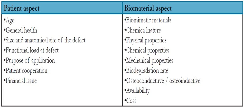
Fig 2: Selection criteria and contributing factors for bone graft application
FUNDAMENTAL CONSIDERATIONS ON BONE SUBSTITUTES
There are four main characteristics considered ideal in bone regeneration (Fig 3)
1. Osteogenesis or osteogenic activity:
Ability of bone formation from viable osteoblasts or preosteoblasts derived from the graft donor area, that are capable of generating cellular proliferation and producing new bone.
2. Osteoconduction:
The capacity of the graft for support or allow cell migration, formation blood vessels and the bone growth in surface.
3. Osteoinductivity:
Refers to the ability of a graft to induce nondifferentiated stem cells or osteoprogenitor cells to differentiate into osteoblasts.
4. Osseointegration:
Refers to contact between the bone and implant without intervening fibrous tissue
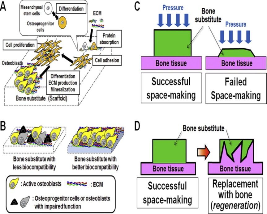
Fig.3: Basic properties of bone substitutes required for bone formation (A)Osteoconductivity. ECM: extracellular matrix. (B) Biocompatibility. (C) Space-making capability. (D) Volume maintenance by replacement with bone over time (regeneration).
It is fundamental that a bone substitute should present at least one of the characteristics described above and only autogenous bone presents them all. Other characteristics considered ideal includes: the remodeling of the bone initially formed in mature lamellar bone, ability to stabilize implants when installed simultaneously to the grafting procedure, low risk of infection, good availability, low antigenicity and physiologically stable, not cause rejection and be ideally be absorbed after the regeneration.10,11
BIOMATERIALS USED FOR BONE REGENERATION IN IMPLANT DENTISTRY:
Autograft:
Autograft is a bone tissue that is separated from one site and implanted in other location in the same individual. They offer a wide pool of growth factors that induce mesenchymal stem cells to differentiate into osteogenic progenitor cells and are subdivided in two groups:
1. Cancellous autografts.
2. Cortical autografts.
Cancellous autografts are retrieved mainly from cancellous bone and upon transplantation, the majority of cells present in the grafts die as result of ischemia. However, the mesenchymal stem cells present in the bone marrow are resistant to ischemia and may survive the grafting procedure.12 The stem cells capacity of survival and proliferation after exposure to changes in the oxygen tension, pH and cytokine environment are the main reason behind the reliability of cancellous bone autograft interventions.
Cortical autografts are segments of cortical bone composed of necrotic bone that provides an osteoconductive support for bone formation, but does not supply significant amounts of living cells. For this reason, revascularization and integration of cortical autografts is slow.12 The main advantage of cortical autografts is the mechanical support that it provides at the graft site.
Autogenous bone transplants are very osteogenic and are the best according to all theoretical bone regeneration requirements. However, there are some disadvantages:
1. Need for second surgical procedure to obtain the graft
2. Donor site morbidity
3. Need for general anaesthesia to obtain extra-oral donor sites
Allografts:
Allograft is defined as tissue that has been harvested from one individual and implanted into another individual of the same species. These are best available alternative to autografts due its similar characteristics. Despite the superior properties of autografts, they are usually preferred by patients as bone grafting material because of the problems associated to donor site surgery in autografts.
Types :
Allografts are obtained from cadaver tissue banks for mineralized freeze-dried (FDBA) or decalcified freeze-dried (DFDBA) bone. Both FDBA and DFDBA are obtained from cortical bone of long bones due to its high content of bone inductive proteins and less antigenic activity than cancellous bone12.
Xenografts:
Xenograft transplants are taken from other species individuals than the recipient. Typically, transplanted materials are natural hydroxyapatite and deorganated bovine bone. These grafts have excellent osteoconductive properties. Xenograft is a bone substitute material derived from deproteinized bone marrow with the hydroxyapatite structure of the highly porous bone similar to cortical bone of human species. The organic component is chemically and thermally treated. These grafts have pore system with a particle size of 0.25-1mm that can be easily invaded by blood vessels resulting in osteoblastic migration the structure consists of a wide interconnection.13 The procedure of processing this may vary and some grafts are treated at high temperature while others are treated at low temperature. Xenogenous transplant treated at low temperature (300ºC) after post transplantation will be fully integrated into human bone tissue, therefore it is called as most physiological transplant.
Limitations:
• Higher risk of recipient’s immunologic reaction
• Possible migration
• Resorption is slow
• Risk of infection
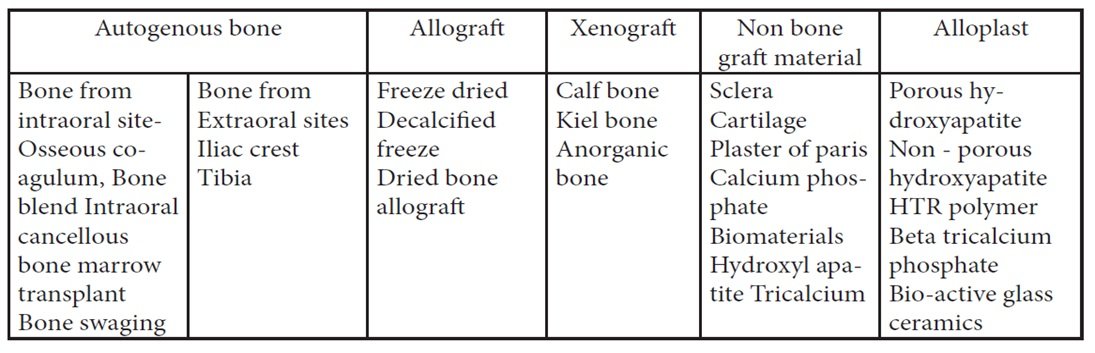
Fig.4: Carranza (1999) classification of bone graft materials14
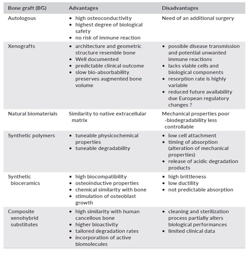
Fig 5: Summary of main advantages and disadvantages of Bone Graft
BONE SUBSTITUTES
Bone substitutes derived from biological products:
Platelet rich plasma (PRP):
Platelet-rich plasma (PRP) is generally used as a gel that is easily obtained with the patient’s blood. Blood is centrifuged through gradient density, and the resulting blood platelets are mixed with thrombin and calcium chloride. Hence, PRP includes an important concentration of platelets and fibrinogen, as well as growth factors such as platelet derived growth factors, vascular endothelial growth factor (VEGF), IGF and TGF. PRP is expected to show pro-coagulant effects due to platelets; however, there is no evidence in the literature of benefits for the addition of PRP to accelerate bone healing.15

Fig 6: Bone substitutes
Platelet Rich Fibrin:
Platelet Rich Fibrin (PRF) is a quite modern platelet concentrate. It is achieved with a simplified preparation, with no biochemical manipulation of blood. This technique does not require anticoagulants or bovine thrombin (or any other gelling agent). This feature makes this product easily usable with a low rate of mistakes during the preparation stage.
Growth factors:
Among various growth factors widely used, BMP (Bone Morphogenic Proteins) requires special mention as Bone morphogenetic proteins (BMPs) are osteoinductive growth factors included in the transforming growth factor β (TGF-β) super family. Genetic engineering allows to synthetize recombinant human BMP (rhBMP-2 and rhBMP-7),16 which can be produced in large quantities and limit risks of contamination.
Hydroxyapatite:
Hydroxyapatite (HA) is part of the apatite family, which are crystalline compounds with crystalline hexagonal lattice. HA has the specific formula (Ca10(PO4)6(OH)2) and is the primary mineral component of teeth and bones.17,18,19. Thus, HA is extremely biocompatible and does not promote an inflammatory response.
Coral:
Corals have interconnected pores and a skeleton quite similar to cortical and spongy bones, and their use as bone substitute has been approved by the FDA in 1992. Coral-based substitutes are mainly calcium carbonate that can be transformed industrially into HA, or they can retain their original state which allows a better resorption by the natural bone.
Bone substitutes derived from synthetic sources:
Ceramics
• Synthetic calcium hydroxyapatite:
Primary inorganic natural bone. The ratio of calcium to phosphorous is 10:6
Tricalcium phosphate:
TCP is chemically similar to HA.20 The ratio of Ca and P is 3:2.
TCP have 2 phases:
• Alfa phase
• Beta phase
When TCP is heated to 900ºC, beta phase is produced which is fully resorbable and is transformed to natural bone after 8-12 months and when heated higher than 1180-degree, alpha is produced (less recommended in clinical practice).
Bioactive glass:
It comprises of calcium salts, phosphate, sodium salts and silicon. The size of pellets varies from 90 – 710 μm with the mean of 300 – 355 μm.21
Calcium carbonate:
Inorganic material which is composed of aragonite (more than 98% CaCO3) which is porous more than 45%. The mean porosity size is about 150 μm.
Composite polymers:
Absorbable
• Synthetic absorbable polymers are well known in medical practice. Many years before suture materials, fixation screws and other products were manufactured from polyacetate and polyglycolic acids.
Non-absorbable:
Non-absorbable composite polymers are the compounds of polymethylmethacrylate (PMMA), polihydroxyethilmethacrylate (PHEMA), small amounts of barium sulphate (roentgenocontrastic) and calcium hydroxide, which directly interact with recipient’s bone, creating CaCO3 appetites.
Nano composite based biodegradable polymer:
The inclusion of nanoparticles into the biopolymer matrix has the dual objective of improving the mechanical properties as well as incorporating Nano topographic features that mimic the nanostructure of natural bone.22
RECENT ADVANCES
Bone tissue engineering:
The complex biology behind the nanostructure and microstructure of bones and their repair mechanisms, which involve various types of chemical and biome chemical signaling amongst different cells, has set strong requirements for biomaterials to be used in bone tissue engineering. Tissue engineering may be used to regenerate bone by combining cells from the body with growth factors and scaffold cells from the body with growth factors and scaffold biomaterials. This combination of cells, signaling molecules and scaffold is often referred to as the tissue engineering triad. Hydrogels are desirable scaffold materials for bone tissue engineering due to their high-water content, cytocompatibility, and mechanical properties to resemble natural tissues.23,24,25. Cell-laden hydrogels have been used for tissue engineering of bone, muscle, cartilage, and other tissues.
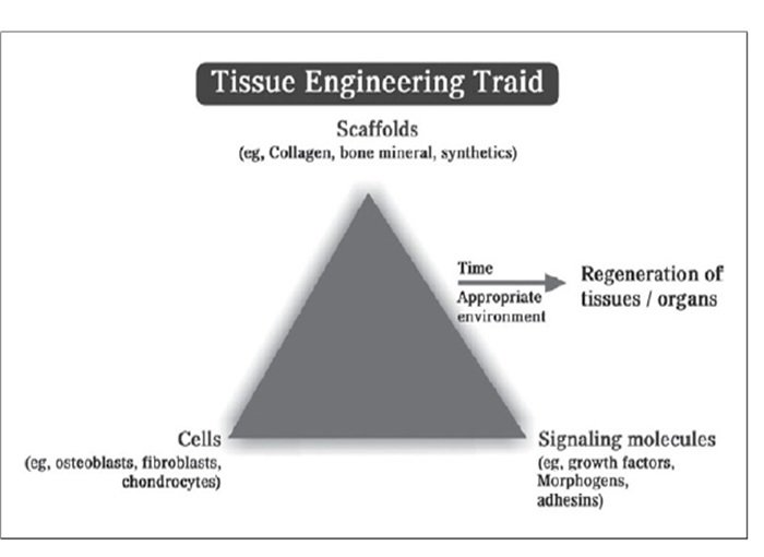
Fig 7: Tissue engineering triad that includes cells, scaffolds, and signalling molecules. This triad is essential for regeneration
Gene therapy
Gene therapy resembles the genetic information transferring to desired target cells, which results in a secure and efficient biological effectiveness leading to the rapid recovery of bone healing. It also adapts for distribution of growth factors in tissue Engineering delivered nucleic acids can induce specific osteogenic growth factors.
Bio – printing /3d printing of bone:
3D printers can print bone tissue tailored to the requirements of the patient and can act as biomimetic scaffolds for bone cell enhancement and tissue growth and differentiation in bone regeneration procedures 3D printed alginate peptide hybrid scaffolds provide a stable environment for growth of stem cells.26 Also composite powders can be printed into scaffolds. Calcium phosphate powders mixed with 3d printing powder based on Calcium sulphate can also be used as bone augmentation material.27
CONCLUSION
This review describes the capabilities and limitations of commercially available bone substitutes based on the insights of biomaterial science from outcomes of clinical and preclinical studies. In summary:
- Autogenous bone is still the gold standard and accelerates initial bone formation to a greater extent than bone substitutes. However, autogenous bone is only effective under favourable recipient conditions and thus requires supplementation with bone substitutes in bone augmentation under severe recipient conditions.
- Currently available bone substitutes induce only osteoconductivity as a scaffold and not osteoinductivity.
- Space-making capability is required for bone augmentation. Particle size, sensitivity to enzymatic or chemical dissolution, and mechanical properties affect the space-making capability of a material.
- The biocompatibility of current bone substitute materials should be improved. Reduced biocompatibility is thought to result from degradation of the extracellular matrix (irregular arrangement of endogenous collagen fibers), generation of micro- or nano- particulates, and excessive release of calcium ions.
- Bioabsorption of remnant bone substitutes and volume maintenance of the augmented tissue over time are inversely related. Bioabsorption during the bone remodeling phase after bone formation is induced by passive chemical dissolution of the material and cellular absorption by osteoclasts, although passive chemical dissolution seems to be predominant.
- Important factors that determine bioabsorption are water solubility and acid resistance, which are determined by chemical composition and the integrity of the apatite structure, respectively.
- At present, no complete bone substitute exists that simultaneously exhibits suitable biocompatibility, bioabsorption, and volume maintenance.
The extensive need for the bone grafts has resulted in the introduction of a number of materials with varied applications. Both natural and synthetic materials have found favourable results in obtaining bone augmentation.
REFERENCES
- Fillingham Y, Jacobs J. Bone grafts and their substitutes. The bone & joint journal. 2016 Jan; 98(1_Supple_A): 6-9.
- Gajavalli S. U, Burugupalli P, Kranthikiran G, Chandrasekharan N. K. Bone Substitutes used in Implant Dentistry. TPDI. July 2018;9(2):32-44
- Axelsson P, Nystrom B, Lindhe J. The long-term effect of a plaque control 600 program on tooth mortality, caries and periodontal disease in adults. Results 601 after 30 years of maintenance. J Clin Periodontol 2004;31:749–57.
- Egusa H, Sonoyama W, Nishimura M, Atsuta I, Akiyama K. Stem cells in 602 dentistry—part I: stem cell sources. J Prosthodont Res 2012;56:151–65.
- Masaki C, Nakamoto T, Mukaibo T, Kondo Y, Hosokawa R. Strategies for 603 alveolar ridge reconstruction and preservation for implant therapy. J 604 Prosthodont Res 2015;59:220–8.
- M. Yamada, H. Egusa, Current bone substitutes for implant dentistry, J Prosthodont Res (2017), http://dx.doi. org/10.1016/j.jpor.2017.08.010
- Haugen H. J, Lyngstadaas S. P, Giuseppe F.R. Bone grafts: which is the ideal biomaterial? J Clin Periodontol. 2019;46(Suppl. 21):92–102.
- Rodriguez R, f Hartmann N, Weingart D. Current concepts of bone regeneration in implant dentistry. J Surg 2015; 10:227-8.
- Ebrahimi M. Bone Grafting Substitutes in Dentistry: General Criteria for Proper Selection and Successful Application.Iosr- Jdms.2017;16(4):75-79
- Kumar J, Jain V, Kishore S, Pal H. Journey of Bone Graft Materials in Periodontal Therapy: A Chronological Review. J Dent Allied Sei. 2016; 5:30-4.
- Cardoso CL, Curra C, Santos PL, Rodrigues MF, Ferreira-Júnior O, de Carvalho PS. Current considerations on bone substitutes in maxillary sinus lifting. Revista Clínica de Periodoncia, Implantología y Rehabilitación Oral. 2016 Aug 1; 9(2):102-7.
- Torres J, Tamimi F, Alkhraisat M, Prados-Frutos JC, Lopez- Cabarcos E. Bone substitutes. InImplant dentistry-the most promising discipline of dentistry 2011. InTech.
- Raghavan R, Shajahan PA, Raj JS, Reini Raju MV, Jishnu S. Review on recent advancements of bone regeneration in dental implantology.International journal of applied dental sciences 2018;4(2):161-163.
- Carranaza FA, Newman MG. Reconstructive Osseous Surgery. In: Clinical Periodontology. Philadelphia, USA: WB Saunders Company. 1999; 8:622-39.
- Andrius Geguzis, Inesa Astramskaite, Dovile Gabseviciute. Bone Substitute Materials in Modern Dentistry. International Annals of Medicine. 2017; 1(4).
- .Teresa M, Kamakshi V. Bone Grafts and Bone Substitutes. Int J Pharm Pharm Sci, Vol 6, Suppl 2, 88-91
- .Kim CS, Kim JI, Kim J, et al. Ectopic bone formation associated with recombinant human bone morphogenetic proteins-2 using absorbable collagen sponge and beta tricalcium phosphate as carriers. Biomaterials 2005;26:2501–2507
- .Engelke WG, Diederichs CG, Jacobs HG, Deckwer I Alveolar reconstruction with splitting osteotomy and microfixation of implants. Int J Oral Ma xillofacImplants. 1997; 12:310-318.
- Roberts TT and Rosenbaum AJ. Bone grafts, bone substitutes and orthobiologics: the bridge between basic science and clinical advancements in fracture healing Organogenesis 2012; 8(4): 114–124.
- Kattimani et al. Hydroxyapatite—Past, Present, and Future in Bone Regeneration. Bone and Tissue Regeneration Insights 2016:7 9–19
- .Saikia KC, Bhattacharya TD, Bhuyan SK, et al. Calcium phosphate ceramics as bone graft substitutes in filling bone tumor defects. Indian J Orthop 2008; 42(2): 169–172.
- Fernandez de Grado G, Keller L, Idoux-Gillet Y, Wagner Q, Musset AM, Benkirane-Jessel N, Bornert F, Offner D. Bone substitutes: a review of their characteristics, clinical use, and perspectives for large bone defects management. sw Journal of tissue engineering. 2018 Jun 2; 9: 1–18.
- Christopherson GT, Nesti LJ. Stem cell applications in military medicine. Stem cell research & therapy. 2011 Dec;2(5):40
- .Anderson JM. The future of biomedical materials. J MaterSci Mater Med 2006;17:1025–1028
- Gurtner GC, Callaghan MJ, Longaker MT. Progress and potential for regenerative medicine. Annu. Rev. Med. 2007 Feb 18;58:299-312.
- Heo EY, Ko NR, Bae MS, Lee SJ, Choi BJ, Kim JH, Kim HK, Park SA, Kwon IK. Novel 3D printed alginate–BFP1 hybrid scaffolds for enhanced bone regeneration. Journal of Industrial and Engineering Chemistry. 2017 Jan 25; 45:61-7
- Sheikh Z, Sima C, Glogauer M. Bone replacement materials and techniques used for achieving vertical alveolar bone augmentation. Materials. 2015 May 27; 8(6):2953-93.
Disclaimer from Guident
The views and opinions expressed in articles published in Guident are solely those of the authors and do not necessarily reflect the views of the editors, the editorial board or the publisher, Ivory India. Guident does not endorse any product, technique or treatment described. Clinical content is intended for qualified dental and medical professionals and is not a substitute for individual clinical judgement. Guident, its editors and the publisher accept no liability for any consequence arising from the use of information published in the journal.
How to cite this article
Dr. Ruchi Kadian, Dr. Jyoti Gupta, Dr. Rahul Nagrath. Bone Substitutes in Implant Dentistry. Guident. 2021 Jan;14(2). Available from: https://guident.in/articles/implantology/bone-substitutes-in-implant-dentistry.html