Esthetic Rehabilitation of Extruded Luxated Maxillary Anterior Teeth
Department of Conservative Dentistry & Endodontics, K.D. Dental College & Hospital, Mathura
Abstract
Traumatic injuries cause multiple kind of injury to dental and periodontal tissues. Extrusive luxation is caused by the action of oblique forces. It is distinguished by the loosening and partial displacement of the tooth out of the socket. This movement is responsible for the almost complete disruption of the periodontal ligament and the rupture of the neurovascular apical bundle. This case report presents a successful esthetic rehabilitation of a patient with avulsion of the maxillary left lateral incisor and extrusion of maxillary left central incisor having grade III mobility.
Introduction
In children and young adults, traumatic dental injuries usually occur, forming 5% of all injuries. 25% of all school children experience dental trauma whereas 33% of adults experienced trauma to the permanent dentition.1 Dentoalveolar traumatic injury resulting in fractured, displaced, or lost anterior teeth have a significant functional, esthetic, speech, and psychological effects on patient’s quality of life.2 Luxations of the permanent teeth are categories as: subluxation, lateral luxation, intrusion and extrusion. Extrusive luxation is a dentoalveolar traumatic injury caused by the action of oblique forces. It is characterised by the loosening and partial displacement of the tooth out of the socket. This transition is responsible for the almost complete disruption of the periodontal ligament and the rupture of the neurovascular apical bundle. Palatal gingival fibres keep the tooth from being avulsed. This traumatic injury can lead to increased tooth mobility and alterations in the pulp or pulp circulation.3,4 This form of injury is relatively common and occurs in 15 to 61% of lesions between the permanent dentition and in 62 to 73% of cases between the lesions involving deciduous teeth.5,6 This present case report presents a case of a patient with avulsion of the maxillary left lateral incisor and extrusion of maxillary left central incisor having grade III mobility.
Case Report
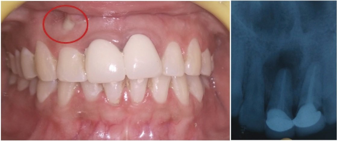
A 18-year-old young female patient reported to the Department of Conservative Dentistry and Endodontics with a chief complaint of trauma to her anterior maxillary region .She reported that she sustained trauma during a fight one day before. Patient presented with history of spontaneous pain in upper front teeth region pain to percussion was present. Clinical examination of maxilla revealed avulsion of the maxillary left lateral incisor and extrusion of maxillary left central incisor with grade III mobility.(Figure-1)
The maxillary right central incisor, maxillary right lateral incisor and maxillary left canine presented with grade 1 tooth mobility. The patient sustained lacerations and mucosal damage in maxillary left anterior region. She had been unable to locate the avulsed teeth.
A periapical radiograph revealed a widened periodontal space around the root of maxillary left central tooth. However, the periapical radiograph showed no evidence of root or bone fracture. After clinical examination, a treatment plan was made. Repositioning the left maxillary central incisor by conventional digital manipulation was planned. A left infraorbital nerve block was given. Careful repositioning of left maxillary central incisor was done by slowly pressing out the teeth along the gingival crevice such that it can be maneuvered apically into its correct position. The tooth was splinted in its normal position by flexible orthodontic wire bonded to labial surfaces by using composite resin for three weeks. (Figure 2-5). The splint was removed after three weeks as the teeth were stabilized. (Figure 6).

Figure 1-Intraoral view after trauma.
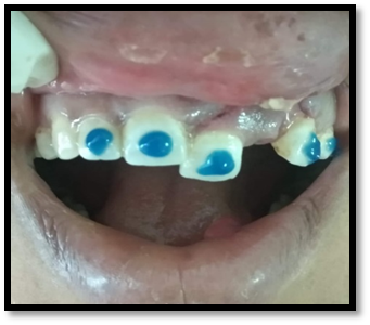
Figure 2- After application of etchant

Figure 3-After application of bonding agent
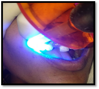
Figure 4-Curing of bonding agent
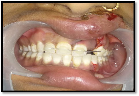
Figure 5-Teeth stabilized with orthodontic wire using composite resin.
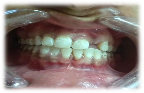
Figure 6- Splint was removed after 3 weeks
Pulp vitality test reported negative results in relation to 11,12,21 and 23. Root canal therapy was initiated in relation to those teeth. (Figure 7-8) Biomechanical preparation was done and calcium hydroxide intracanal medicament was placed in the root canals for 7 days. Thereafter, obturation of the root canals was carried out followed by placement of permanent restoration in the respective teeth. Prosthetic rehabilitation was done in relation to 11,12,21,22 and 23. (Figure 9-11) Unfortunately, due to pandemic Covid-19 lockdown, patient got her prosthetic restoration done from some private practitioner.

Figure 7- Working length irt 11,12,21,23.
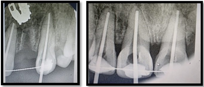
Figure 8- Master cone irt 11,12,21,23.

Figure 9- crown cutting was done irt to 11,12,21,23.

Figure 10-Polyvinyl siloxane impression was made.

Figure 11-Prosthetic rehabilitation irt to 11,12,21,22,23.
Discussion
From an endodontic perspective, pulp necrosis with inflammation, pulp canal calcification, ankylosis and root resorption are the most complicating factors for luxation cases. Factors affecting the prognosis of luxated teeth are the degree of displacement, treatment time delay, root maturation and concomitant crown fractures. In most of the cases, within four months’ pulp necrosis in luxated teeth become quite evident. Root resorption frequently occurs within the first five months of injury and can progress very rapidly, particularly in immature teeth. Thus, frequent follow-up examination of such cases is recommended. In fact, long-term follow-up is required as in some cases pulp necrosis may appear at a much later date.7
The present case report, presents a case of a patient with extruded maxillary left central incisor. In such case of extrusive luxation, the tooth is extruded from its socket so that minimal damage to the socket wall occurs. A recent study involving a paediatric population showed that extrusive luxation led to pulp necrosis in 43 % of teeth, usually within a year. Pulp canal calcification was noted in 35 % and progressive root resorption in 5.5 % of cases. A direct association between the degree of extrusion and the occurrence of pulp canal obliteration, but not necrosis, has been identified in this study.8
The ideal treatment for this type of trauma comprised of thorough clinical and radiographic evaluation, repositioning of the tooth in its socket with digital pressure followed by semi-rigid fixation of the affected tooth to surrounding teeth for 2 weeks. Occlusal adjustment and orientations for oral hygiene, prognosis, and a follow-up is recommended.4,9,10
Therefore, in our case, the tooth was splinted in its correct position by flexible orthodontic wire bonded to labial surfaces by using composite resin for three weeks. Proper oral hygiene instructions were given and follow up of the case was done. Patient presented asymptomatic after one year without any clinical signs of mobility and displacement of the teeth.
Conclusion
Traumatic dentoalveolar injuries may cause functional, esthetic, and psychological problems to the patients. Such cases often require a multidisciplinary treatment approach. This case report highlight the successful esthetic rehabilitation of extruded luxated maxillary left central incisor. The patient was asymptomatic after one year of treatment with no external or internal root resorptions.
References
- International Association of Dental Traumatology.Dental Trauma Guidelines.Revised 2012.https://www.iadt-dentaltrauma.org/1-9%20%20iadt%20guidelines%20combined%20-%20lr%20-%2011-5-2013.pdf(Last accessed on 11february 2021)
- Cortes MI, Marcenes W, Sheiham A. Impact of traumatic injuries to the permanent teeth on the oral health-related quality of life in 12-14-year-old children. Community Dent Oral Epidemiol 2002; 30:193–8.
- Andreasen JO, Andreasen FM, Bakland LK, Flores MT. Traumatic dental injuries – the guide. Oxford: Blackwell Munksgaard; 2003.
- Andreasen JO, Andreasen FM, Andersson L. Textbook and Color Atlas of Traumatic Injuries to the Teeth. 4th ed. Oxford, England, Wiley-Blackwell: 2007.
- Glendor U, Halling A, Andersson L, Eilert-Petersson E. Incidence of traumatic tooth injuries in children and adolescents in the county of Västmanland, Sweden. Swed Dent J 1996; 20:15-28.
- Haavikko K, Rantanen L. A follow-up study of injuries to permanent and primary teeth in children. Proc Finn Dent Soc 1976; 72:152-162.
- Andreasen FM. Pulpal healing following acute dental trauma: clinical and radiographic review. Pract Proced Aesthet Dent 2001; 13:315-322; quiz 324.
- Lee R, Barrett EJ, Kenny DJ. Clinical outcomes for permanent incisor luxations in a pediatric population. II. Extrusions. Dent Traumatol 2003; 19:274-279.
- Araújo, MA.; Valera, MC. Injuries by luxation. In: Araujo, MA.; Valera, MC., editors. Clinical treatment of dental trauma. 1st ed. São Paulo: Artes Médicas; 1999. p. 187-196.
- Dumsha TC. Luxation injuries. Dent Clin North Am 1995; 39:79-91.
Disclaimer from Guident
The views and opinions expressed in articles published in Guident are solely those of the authors and do not necessarily reflect the views of the editors, the editorial board or the publisher, Ivory India. Guident does not endorse any product, technique or treatment described. Clinical content is intended for qualified dental and medical professionals and is not a substitute for individual clinical judgement. Guident, its editors and the publisher accept no liability for any consequence arising from the use of information published in the journal.
How to cite this article
Dr. Deepika, Dr. Muhammad Mutiur Rahman, Dr. Ajay Nagpal. Esthetic Rehabilitation of Extruded Luxated Maxillary Anterior Teeth. Guident. 2021 Jan;14(2). Available from: https://guident.in/articles/endodontics/esthetic-rehabilitation-of-extruded-luxated-maxillary-anterior-teeth.html