Dental Complications After COVID-19: Understanding the Pathophysiology: Part-5
Wisdom Dental Clinics, Jaipur, India
ABSTRACT
Severe covid-19 cases are currently being managed with systemic drugs. Dentists are frequently being reported with cases presenting rare conditions which are destroying jawbones after covid -19 infection.Opportunistic infection have become concern for such individuals.Here we report a case with rare condition affecting the jaw presenting with similar condition in our previous case and the questionable underlying etiology with description of pathophysiology and diagnosis related to such diseases followed with conservative surgical intervention removing sequestrum only.Surgeons should be aware of secondary invasive conditions in post covid-19 infected individuals.
Keywords: Covid-19, fungal infection, secondary infection, questionable, etiopathogenesis
INTRODUCTION
While the world is battling against covid-19 sepsis has outraged a severe condition. Oral surgeons are repeatedly encountered with cases presenting rare fungal infections, osteomyelitis, thromboembolic event and MRONJ/CRONJ related to jaws in covid-19 individuals undergoing resection of affected portion of jaw.covid-19 and its treatment against these kind of backdrop has become the major issue for the disaster. We have come across different cases in past few months dealing with almost similar signs and symptoms and presenting with deleterious condition related to maxilla. Meanwhile second wave of covid-19 has come still such cases are repeatedly found.
Around 77 million cases in population of India diabetes is one of the fastest growing known epidemic.one of the cross sectional study from all the states of India, revealed 47% of people are not aware of their diabetic status and only quarter of patients have yet achieved their glycemic control upon treatment.1
The dreadful combination of diabetes and SARS-CoV-2 has been repeatedly noted in various studies around the world.2
Mucorales are widespread moulds, found abundantly in the surroundings feeding on decaying organic matter various number of studies have revealed huge mould spore counts in hospital air which is due to hot,humid climatic condition.3 Mucormycosis is a rare opportunistic infection mainly caused by saprophytic fungi group,which belongs to mucorales and zygomycetes class.4
These organisms have huge tendency to invade vascular system,thus forming thrombi,leading to low blood supply and finally causing tissue necrosis.Generally ,the condition may present by involving Rhinocerebral,pulmonary,gastrointestinal system ,cutaneous and disseminated forms.5,6,7
Maxilla is the primary bone of the face that is vital structure of viscerocranium. Maxillary necrosis occurs rarely as compared to the necrosis of mandible due to its own high vascular supply.8
In our previous cases we suspected for osteomyelitis, thromboembolic event, MRONJ/CRONJ, mucormycosis associated with diabetes interlinked with post covid-19 infection. Here we present with a case with similar condition as in our previous suspected case of mucormycosis with multiple questionable etiopathogenesis.
CASE REPORT
After presenting our four parts and respective cases dealing with covid-19 dental complication series of cases we here by move to our last case of series, in this case too we were reported with a male patient presenting with almost very similar signs and symptoms as shown in our previous case except that here patient did not present with pus discharge. After taking the history it was revealed that patient was operated by ENT surgeon where his maxillary sinus was drained and cleaned i.r.t tooth no 6,7,8 in right maxillary arch and patient was under antifungal therapy of amphotericin b and itraconazole for 1 month still could not recover so he visited our clinic.
Upon intraoral examination we found oedematous and inflammed gingiva in right maxillary arch.(Figure.1 a,b)
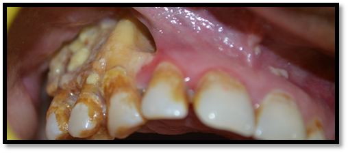
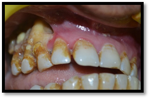
Figure 1 a,b: Showing inflammed gingiva with infection in right maxillary arch
Patient presented with few investigations & OPG which revealed bone loss and radiolucency i.r.t 6,7,8 in right maxillary arch. (Figure.2)
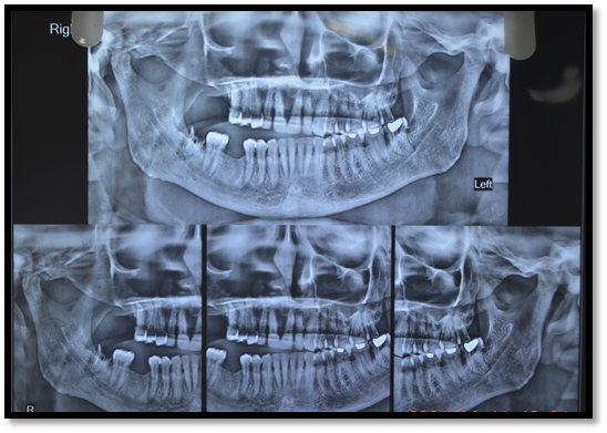
Figure 2: Bone loss and radiolucency in right maxillary arch
We further went for CBCT to rule out soft and hard tissue infection of the concerned area and looking for any spread of infection. Showing absence of right maxillary sinus as well as alveolar bone till distal aspect of premolar,with left maxillary arch osteomyellits? (Figure.3a,b,c)
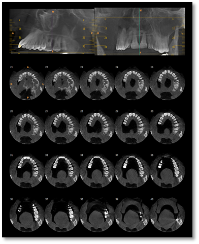
Figure 3a: Axial view of maxilla
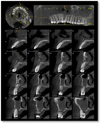
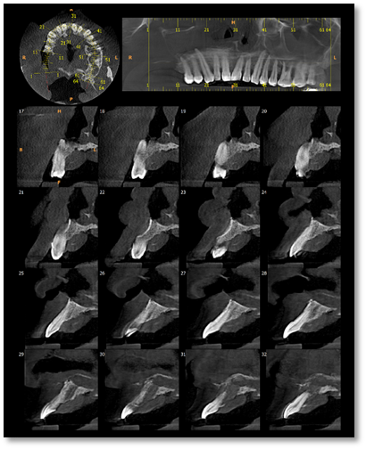
Figure 3b,c: CBCT image of maxillary arch
Continuing further with our case we went for CBC and random blood glucose levels to look for comorbid condition .Report showed Hb 11.90gm/dl (low),raised TLC count and neutrophil count.Lymphocyte 15.40% (low) with decreased values of PCV and MCHC. Random blood glucose level within range. (Figure.4)
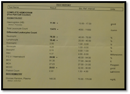
Figure 4: Report of CBC and random blood glucose level
To rule out any infection and inflammation and blood clotting disorder we thought to go for BT,CT and CRP which revealed increased value of CRP showing some underlined infection.(Figure.5)
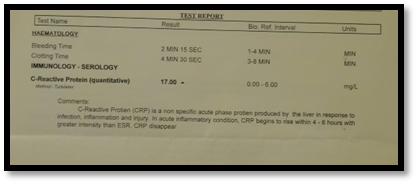
Figure 5: Increased value of CRP 17 mg/L
To find out any other systemic condition patient was asked to go for liver function test which showed increased in values of serum alkaline phosphatase and protein levels.(Figure.6)
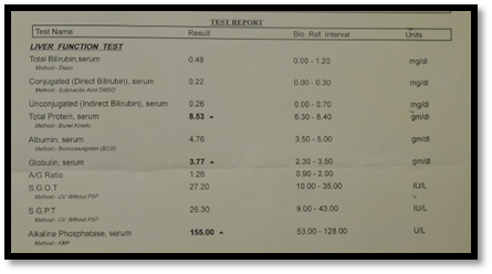
Figure 6: Liver Function Test report
Looking at the clinical condition of the case so ,for evaluating any underlying thromboembolic event related to this condition we continued to consider for D-dimer test values which came to be within range.(Figure.7)
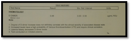
Figure 7: D-dimer 0.48 ug/mL
We almost went for most of the tests and radiographs still couldnot get the outcome for the root cause of the condition ,so we thought it could be some fungal or malignant condition therefore,we went for biopsy which gave overall morphology of subacute osteomyelitis, inflammatory granulation tissue with no malignancy or fungus evidence.(Figure.8)
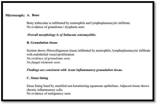
Figure 8: Biopsy report showing no evidence of fungus or malignancy
After clinically evaluating and going through various test findings the root cause of the condition still could not be located.To prevent further damage from the infection we thought to go for surgical intervention to remove the necrotic area in order to preserve the near by hard and soft tissues followed with antibiotic therapy. Same treatment protocol was followed as done with previous patients under local anesthesia.Upon regular recall dental visits patient responded well with the healing of affected area.
DISCUSSION
Further going on to our last case of covid -19 dental complication series of cases here also we were reported with a male patient showing with almost similar signs and symptoms as presented in our earlier case of mucormycosis except that in this present case of ours there is no pus discharge from the affected area.In this part we discuss about the multiple questionable etiopathognesis linked with the condition which can be one of the reason for such encountered disease.
We were frequently reported with cases presenting with similar conditions affecting only maxilla reported with male gender in common in past few months meanwhile second wave hit the country dreadly still such similar cases are repeated we thought that multiple reasons could be one of the etiology leading to pathogenesis of such conditions. We had a notion that it could be a retrograde infection either through sinuses (like sphenoid, frontal, maxillary etc) & then involving maxilla and finally reaching to maxillary alveolus.
In all of our cases presented we found attached gingiva to be intact.
The following questions aroused which could be one of the responsible etiologic pathogenesis:
1. Due to primary infection?

Necrosis if found present in maxilla it is due to either bacterial invasion causing osteomyelitis or due to viral infection like herpes or lastly due to any trauma or invasion by fungus leading to mucormycosis.9
2. Due to secondary infection?

All the previously reported cases were of secondary infection.
Various secondary etiologic factors can lead to secondary infection like underlying comorbid condition, immunomodulatory therapy by steroids and tocilizumab in covid-19 subjects:
Tocilizumab is known recombinant humanized monoclonal antibody which is used against IL-6 i.e Interleukin-6 receptor. It effectively reduce the IL-6 levels in covid-19 individuals.10 Individuals treated with tocilizumab had a higher risk of developing serious bacterial ,skin as well as soft tissue infection.11-12
Steroids do form one of the cornerstone in covid-19 treatment therapy.Steroids do modulate the inflammatory response as a standard of care recognised in WHO guidelines.13 IL -6 also promotes for the viral and bacterial clearance,thus suppressing it with immunomodulating drugs which could lead to secondary infection.Warnings about the high risk involved with the use of immunosuppresants have become more numerous in last year causing bacterial and dreadful fungal infections.14
Most of the covid-19 infected individuals are found reported with lymphopenia in whom use of steroids increases the risk for fungal infections like aspergillosis.15 Our case also presented with lower lymphocyte count than normal.
In one of the studies which described about a 60yr old male patient developed with rhino-orbital mucormycosis,suggested that very extensive use of steroids /monoclonal antibodies/broad spectrum antibiotics may develop or in return exacerbate pre-existing fungal disease.16
Mucormycosis many a times appear as the diabetes-defining illness, it still remains one of the most devastating complications in uncontrolled diabetics having mortality rates ranging between 40-80%. India contributes to nearly about 40% of the global burden of this “rare mould” infection as it is called in western literature, with an estimated prevalence of nearly 140 cases per million population.17 High level of glucose lead to altered phagocytic activity of granulocytes and in diabetic ketoacidosis there is high risk due to increased serum iron levels.
Post covid-19 infection is what that occurs after SARS-CoV-2 has already had a rampage.It lead to imperfectly regulated innate immunune response,ciliary dysfunction,cytokine storm,thrombo inflammation,microvascular coagulation and finally leading to an eventual immune system exhaustion.
These events cascade down leading to facilitate secondary bacterial and fungal infections more in critically ill subjects. Further,the use of corticosteroids and anti IL- 6 stategies in these more susceptible individuals along with higher number of fungal spore counts in the surrounding environment create the ideal settings for mould infections.The coronavirus might be found associated with wide range of disease patterns which is ranging from mild –moderate to severe pneumonia.A wide range of bacterial as well as fungal co infections are found to be associated with pre-existing comorbidities like diabetes, lung disease and low innate immunity.
Pathophysiology of mucormycosis can be represented as follows:

With our knowledge we looked for all the possible causes relating to this condition we went through number of tests including radiological , pathological, haematological and histological to evaluate the root cause we even thought to look for any underlying thromboembolic event so went for D-dimer values but could not relate for the cause as its value was within reference range even histology did not gave any evidence of fungus or malignancy.
To make the diagnosis we must do all the investigations simultaneously:
- Radiological
- CT PNS
- MRI brain with orbit and PNS
- Nasal tissue for KOH mount
- Histology
- Blood and other related investigations
Still question arises why it is only involving maxilla why not mandible? Is it due to presence of maxillary sinus?
Another question aroused while going through this case was as fungal infection of mucormycosis is very destructive infection why is it not spreading to mandible ,why is it just limited to maxilla in case of oral cavity?
Though maxilla is rarely involved still factors which are predisposing to osteomyelitis include dental infections, maxillary sinusitis, trauma, and radiation. The two main causes of maxillary involvement are dental infections and sinusitis.18
Moving further we got into another questionable situation if it is due to covid-19 and is secondary fungal infection then why it is also that Some patients are not diagnosed by RT-PCR & no antibodies are also there?? Why did we still got healing results though we did not used any antifungal therapy in this present case? We followed with antibiotic therapy only .It was also found to be related with 3M and diabetes i.e Men, Maxilla and Mucormycosis.
CONCLUSION
Covid-19 is associated with secondary infection which might be bacterial or fungal due to dysregulation of innate immunity. Additionally, extensive use of steroids/broad spectrum antibiotics as a basic part of armamentarium to treat covid-19 may lead to exacerbation of any pre existing fungal disease. Clinicians should be aware of possibility of invading secondary infections with pre existing higher risk factors for early and prompt diagnosis with reduction in morbidity and mortality rate. Use of therapeutic drugs should be monitored at low dose with short duration of time.Conservative surgical approach should be followed removing only sequestrum part. More studies are needed to know the exact etiopathogenesis of such conditions involving only maxilla with male predominance and know its possible course of action, is it the same infection invading other body parts? There should be multi disciplinary approach of diagnosis in post covid-19 patients.
REFERENCES
- Prenissl J, Jaacks LM, Mohan V,et al. Variation in health system performance for managing diabetes among states in India: a cross-sectional study of individuals aged 15 to 49 years. BMC Med 2019; 17:92.
- Lim S, Bae JH, Kwon HS,et al. COVID-19 and diabetes mellitus: from pathophysiology to clinical management. Nat Rev Endocrinol 2021; 17:11–30.
- Rudramurthy SM, Singh G, Hallur V et al. High fungal spore burden with predominance of Aspergillus in hospital air of a tertiary care hospital in Chandigarh. Indian J Med Microbiol2016; 34:529-532.
- Paulltauf A. Mycosis mucorina. Virchows Arch Pathol Anat. 1885;102:543–64. [Google Scholar]
- Gonzalez CE, Couriel DR, Walsh TJ. Disseminated zygomycosis in a neutropenic patient: Successful treatment with amphotericin B lipid complex and granulocyte colony-stimulating factor. Clin Infect Dis. 1997;24:192–6. [PubMed] [Google Scholar]
- Cuvelier I, Vogelaers D, Peleman R, Benoit D, Van Marck V, Offner F, et al. Two cases of disseminated mucormycosis in patients with hematological malignancies and literature review. Eur J Clin Microbiol Infect Dis. 1998;17:859–63. [PubMed] [Google Scholar]
- Pagano L, Ricci P, Tonso A, Nosari A, Cudillo L, Montillo M, et al. Mucormycosis in patients with haematological malignancies: A retrospective clinical study of 37 cases. GIMEMA Infection Program (Gruppo Italiano Malattie Ematologiche Maligne dell’Adulto) Br J Haematol. 1997;99:331–6. [PubMed] [Google Scholar]
- A. Urs, H. Singh, S. Mohanty, and P. Sharma, “Fungal osteomyelitis of maxillofacial bones: rare presentation,” Journal of Oral and Maxillofacial Pathology, vol. 20, no. 3, p. 546, 2016. View at: Publisher Site | Google Scholar
- J. A. Kumar, P. Babu, K. Prabu, and P. Kumar, “Mucormycosis in maxilla: rehabilitation of facial defects using interim removable prostheses: a clinical case report,” Journal of Pharmacy and Bioallied Sciences, vol. 5, no. 2, p. S163, 2013. View at: Publisher Site | Google Scholar
- Sheppard M, Laskou F, Stapleton PP, Hadavi S, Dasgupta B. Tocilizumab(Actemra). Hum Vaccin Immunother. 2017 Sep 2;13(9):1972-1988. doi:10.1080/21645515.2017.1316909. PMID: 28841363; PMCID: PMC5612212
- Narazaki M, Kishimoto T. The Two-Faced Cytokine IL-6 in Host Defense and Diseases. Int J Mol Sci 2018;19.
- Davis JS, Ferreira D, Paige E, Gedye C, Boyle M. Infectious complications of biological and small molecule targeted immunomodulatory therapies. Clin Microbiol Rev. (2020) 33:e00035-19. 10.1128/CMR.00035-19
- Lamontagne F., Agoritsas T., Macdonald H., Leo Y.S., Diaz J., Agarwal A. A living WHO guideline on drugs for COVID-19. BMJ. 2020 Sep 4;370:m3379. doi:10.1136/bmj.m3379
- Kimmig L.M., Wu D., Gold M., Pettit N.N., Pitrak D., Mueller J. IL6 inhibition in critically ill COVID-19 patients is associated with increased secondary infections. medRxiv. [Preprint] 2020 Sep 12 doi: 10.1101/2020.05.15.20103531
- Deana C, Vetrugno L, Bassi F, De Monte A. Tocilizumab administration in COVID-19 patients: Water on the fire or gasoline? Med Mycol Case Rep. 2021 Mar;31:32-34. doi: 10.1016/j.mmcr.2021.01.002. Epub 2021 Jan 21. PMID:33520634; PMCID: PMC7826035
- Mehta S, Pandey A. Rhino-Orbital Mucormycosis Associated With COVID-19.Cureus. 2020 Sep 30;12(9):e10726. doi: 10.7759/cureus.10726. PMID: 33145132;PMCID: PMC7599039
- Prakash H, Chakrabarti A. Global Epidemiology of Mucormycosis. J Fungi (Basel) 2019; 5:26.
- Peravali R. K., Jayade B., Joshi A., Shirganvi M., Bhasker Rao C., Gopalkrishnan K. Osteomyelitis of maxilla in poorly controlled diabetics in a rural Indian population. Journal of Maxillofacial and Oral Surgery. 2012;11(1):57–66. doi: 10.1007/s12663-011-0283-0. [PMC free article] [PubMed] [CrossRef] [Google Scholar]
Disclaimer from Guident
The views and opinions expressed in articles published in Guident are solely those of the authors and do not necessarily reflect the views of the editors, the editorial board or the publisher, Ivory India. Guident does not endorse any product, technique or treatment described. Clinical content is intended for qualified dental and medical professionals and is not a substitute for individual clinical judgement. Guident, its editors and the publisher accept no liability for any consequence arising from the use of information published in the journal.
How to cite this article
Dr. Gaurav Gupta, Dr. D.K. Gupta, Dr. Neelja Gupta. Dental Complications After COVID-19: Understanding the Pathophysiology: Part-5. Guident. 2021 Jul;14(8). Available from: https://guident.in/articles/general/dental-complications-after-covid-19-understanding-the-pathophysiology-part-5.html