
Root End Resection: A Case Series
Department of Conservative Dentistry and Endodontics, Seema Dental College And Hospital, Rishikesh, U.K, (Professor & HOD.), Department of Conservative Dentistry and Endodontics, D.J. College of Dental Sciences & Research, Modinagar, U.P
ABSTRACT
Traumatic dental injuries (TDIs), especially crown fractures and luxations, are prevalent in children and young adults affecting permanent teeth. Successful outcomes hinge on accurate diagnosis, meticulous treatment planning, and consistent follow-up. This article details a series of case reports on the surgical management of such traumatic injuries that led to periapical lesions, utilizing root end resection with specialized root end filling materials.
INTRODUCTION
Apical surgery, which involves root end resection, aims to prevent bacteria from leaking from the root canal system into the surrounding periradicular tissue.
According to Lieblich, periapical surgery should only be considered for teeth that show no signs of fracture and have good periodontal health. This means less than 25% vertical bone loss and periodontal pockets smaller than 5 mm. Additionally, the tooth must have enough structure remaining to support a prosthesis, and the patient needs to be healthy enough to undergo the procedure.
The European Society of Endodontology (ESE) updated its guidelines for apical surgery indications in 2006. These now include:
Indications for Apical Surgery (ESE 2006)
- Apical periodontitis (identified through X-rays or symptoms) linked to an obstructed root canal. This applies when the obstruction can't be removed, repositioning it isn't practical, or attempting to do so carries a high risk of damage.
- Extruded root canal material accompanied by clinical or radiological evidence of apical periodontitis or persistent symptoms over an extended period1-5.
Contraindications for apical surgery include inadequate periodontal health, tooth with vertical root fracture, compromised medical history such as blood dyscariasis, uncontrolled diabetes.
CASE REPORTS
Case Report 1
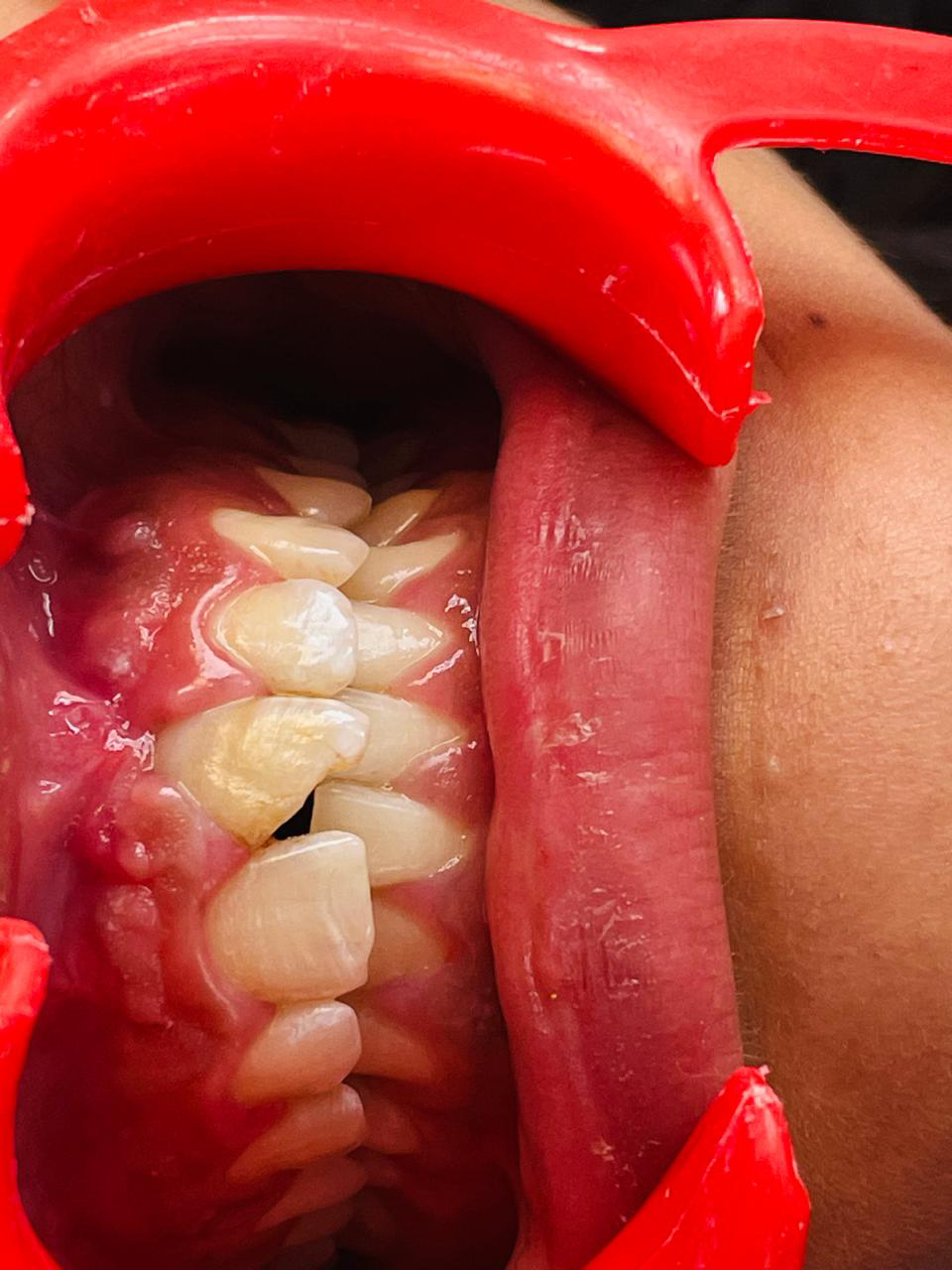
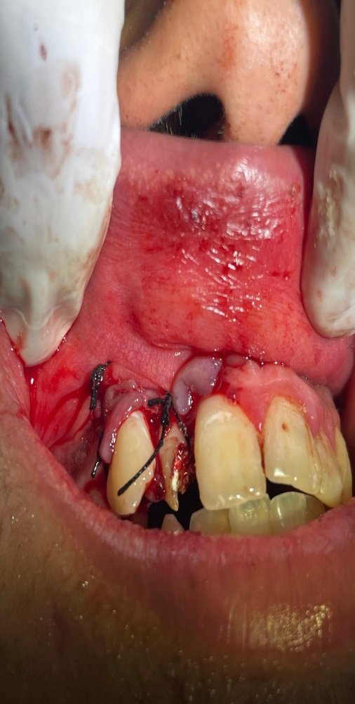
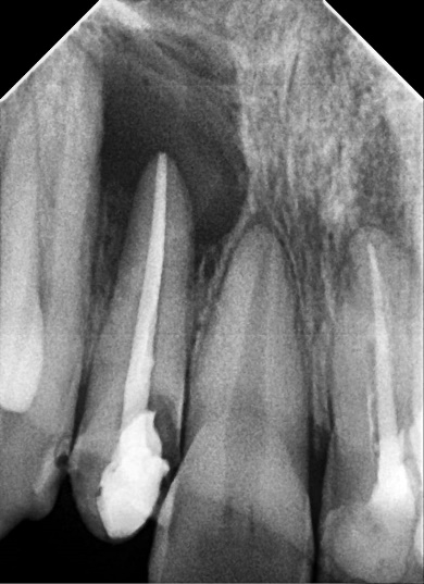
A patient came to the department with the chief complaint of fractured tooth & mobility of teeth in upper front tooth region for 6 month. On taking the history, it was revealed that patient had met with an accident during her childhood. On examination grade 2 mobility was seen. Radiograph revealed a large periapical lesion involving 21 and 22. Treatment plan was made of root canal therapy w.r.t 21, 22 followed by root end resection w.r.t 21,22 Prior to surgery, hematological investigations were carried outstand the patient signed written consent.

Preoperative image
post operative image

Post operative radiograph
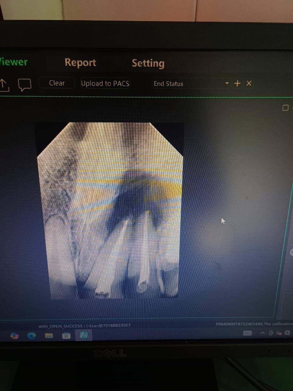
Preoperative radiograph

3 months follow up
Case Report 2
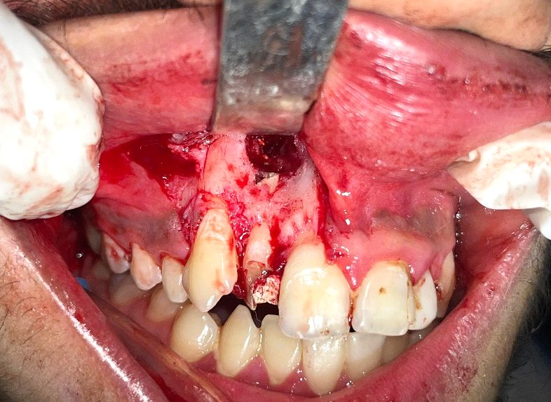
A 32-year-old male came to the Department of Conservative Dentistry and Endodontics (Seema Dental College and Hospital) with the chief complaint of pain and pus discharge for 1 week. Patient gave history of trauma 3 years back while playing football. It was seen that the root canal treatment w.r.t 12 was done by the previous dentist. Patient gave no history of any systemic illness. On examination, there was no obvious swelling. On radiographic examination periapical radiolucency involving 12 can be well appreciated. Diagnosis made was Ellis class III fracture. Treatment plan made was root canal treatment followed by root end resection.
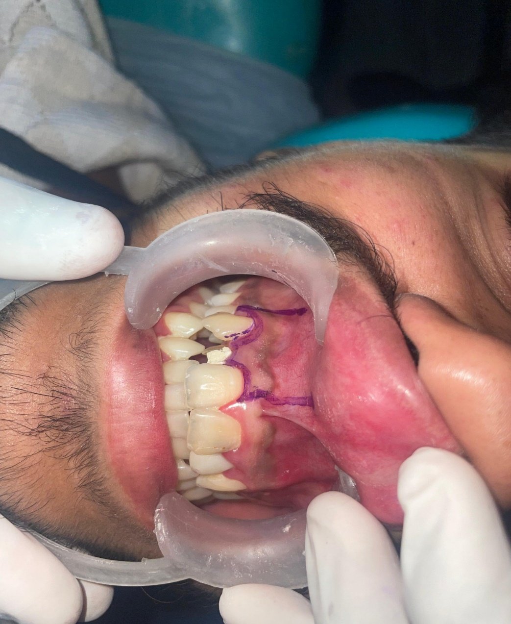
Preoperative radiograph

Post operative radiograph

3 months follow up
Discussion
Periradicular surgery aims to access the affected area, assess root anatomy, and place a biocompatible root-end filling to promote tissue regeneration. Flap design, like the full-thickness or Ochsenbein-Luebke, must ensure surgical access and adequate vascularization. During the procedure, bone sounding helps locate the diseased area, and removing 3mm of the root apex eliminates accessory canals contributing to disease. A 3mm deep, Class I cavity is then prepared using ultrasonic tips, creating a less smudged surface for the Mineral Trioxide Aggregate (MTA) filling. After careful cleaning and irrigation, the flap is repositioned and compressed, followed by suturing with 3-0 silk. Post-operative ice packs minimize clotting and enhance healing. Follow-up at three and six months showed evidence of healthy bone formation and reduced periapical radiolucency6.
Conclusion
Apical surgery is now a reliable option for treating teeth with apical pathology untreatable by conventional endodontics. A high-quality apical seal, achieved with materials like Mineral Trioxide Aggregate (MTA), is key to successful treatment. This is particularly relevant for teeth with persistent periapical radiolucencies and necrotic pulps.
References:
- Serrano-Giménez M, Sánchez-Torres A, Gay-Escoda C. Prognostic factors on periapical surgery: A systematic review. Med Oral Patol Oral Cir Bucal. 2015;20(6):715–22. doi:10.4317/medoral.20613.
- von Arx T. Apical surgery: A review of current techniques and outcome. doi:10.1016/j.sdentj.2010.10.004. Saudi Dent J. 2011;23(1):9–15.
- Franco PB, Karlis V. Apicoectomy in atlas of oral and maxillofacial surgery. Kademani D, Tiwana P, editors. St. Louis, MO: Elsevier; 2016.
- Ajayi J. Comparative study of treatment outcome in apicectomies with or without root-end filling. Ann Ibadan Postgraduate Med. 2018;16(2):109–14.
- Wang N, Knight K, Dao T, Friedman S. outcome in endodontics: I and II: apical surgery. the Toronto Treatment Study. Phases J Endod. 2004;30(11):751–61. doi:10.1097/01.don.0000137633.30679.74.
- Frank CS, Sweta BS, Meetu RK, Bekir K, Syngcuk K. Outcome of endodontic surgery: a meta-analysis of the literature- part I: comparison of traditional root end surgery and endodontic microsurgery. JOE. 2010;36(11):1757–65.
Disclaimer from Guident
The views and opinions expressed in articles published in Guident are solely those of the authors and do not necessarily reflect the views of the editors, the editorial board or the publisher, Ivory India. Guident does not endorse any product, technique or treatment described. Clinical content is intended for qualified dental and medical professionals and is not a substitute for individual clinical judgement. Guident, its editors and the publisher accept no liability for any consequence arising from the use of information published in the journal.
How to cite this article
Dr. Shalvini Kancha, Dr. Shubham Dalvi, Dr. Anil Dhingra Guide. Root End Resection: A Case Series. Guident. 2025 Oct;18(11). Available from: https://guident.in/articles/endodontics/root-end-resection-a-case-series.html


