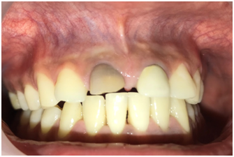
Magnification-Loupes VS Dental Operating Microscopes
Upskill Dental Academy Mumbai, India
The biggest problem most dentists face is the decision to invest in magnification and recovery on investment .In my previous article i have discussed how the role of magnification in dentistry has risen with advancement in technology.
Although microscope training was made mandatory in the year 1997(ADA)1,practioners are still facing a dilemma on making a choice between loupes and dental microscope .This article reviews the benefits and disadvantages of both dental microscopes as well as dental loupes.
Dentists face many challenges as they work in a small, inaccessible, wet, and dark environment. These key physical elements must be controlled once we have diagnosed a patient’s problem and begin treatment. The solutions to these challenges involve proper isolation, magnification, and illumination. The use of a remote light source carried via fiberoptic cabling to a lens that is either attached to a headband or to bioptic magnifying glasses(loupes) is the first choice of most of the practioners.2
Although magnification improves the accuracy of clinical and diagnostic skills, it requires an understanding of optical principles that govern all magnification systems. The assumption that “more magnification is better” must always be weighed against the decrease in field of view and depth of focus that can occur as magnification increases, which is a problem more common with dental loupes than operating microscopes
Loupes are fundamentally dual monocular telescopes with side-by-side lenses convergent to focus on the operative field. The magnified image formed has stereoscopic properties by virtue of their convergence.
Although dental loupes are widely used ,they being economical and easy to use, they have disadvantages compared with the microscope. The clinician's eyes must converge to view the operative field. This can result in eyestrain, fatigue, and even pathologic vision changes, especially after prolonged use.
Another disadvantage of simple loupes is that they are greatly affected by spherical and chromatic aberration. This distorts the image shape and color of objects being viewed.
Dental loupes provide a limited range of magnification, 1.5× to 6×. Loupes delivering magnification of less than 3× are usually inadequate for the visual acuity necessary for precision work. Surgical loupes providing magnification of more than 4× are impractical because of their small field of view, shallow depth of focus, and excessive weight. Excessively heavy loupes can make it difficult to maintain a stable visual field. Although the initial expenses involved in procuring a pair of dental loupes is much lesser than a dental microscope ,it falls short in several aspects both for long term use as well as high magnification to name a few.
When dentists use loupes consistently (that is, they use their loupes all the time, not just for inspection) and want to go above 3x in power, they are ready to move up to a dental microscope. One of the great attributes of the dental microscope is that it has multiple levels of magnification (five steps) and a built-in coaxial, high-intensity light source.3
The optical elements of surgical microscopes are more advanced than those in loupes, depth-of-focus and field-of-view characteristics are greatly enhanced. Surgical microscopes have objective lenses with various working distances. A useful range in dentistry is 250 to 350 mm.
Their manoeuvrability provides visual access to every area of the mouth and is an important factor in choosing to use a surgical microscope.

Fig 1_dental loupes
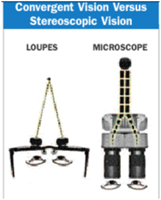
Fig 2_pic courtesy -Magnification alternatives -Seeing is believing, Glenn A. van As, BSc, DMD

Fig 3
The surgical microscope offers greater versatility than dental loupes, by providing a range of magnification with superior optical performance. A surgical microscope can last an entire career, making its long-term expense practical. Proficient use of the microscope requires training and practice. Surgical microscopes designed for dentistry employ Galilean optics, which have binocular eyepieces joined by offset prisms, to establish a parallel optical axis and permit stereoscopic vision without eye convergence or eyestrain. An additional binocular eyepiece can aid the microsurgical assistant.
Documentation is important for patient and professional education and for dental–legal reasons. The surgical operating microscope is an ideal platform for documenting pathology and clinical procedures. Digital images can be captured using a beam splitter and camera attachment. An inbuilt camera permits the operator to record, all the procedure , without interrupting the procedure. These images represent the operating field exactly as the operator sees it, as opposed to a camera view over the shoulder. High-definition video cameras capture both still and video images simultaneously to permit documentation of procedures for educational purposes.4
Hence the use of magnification is always a slow process of graduating from lower magnification to higher magnification. This happens only when the operator realises the benefits of the same and desires to graduate to the use of higher magnification. Proficiency in working with dental loupes and/or dental microscopes is a slow process and it involves training from an experienced mentor,trials/lookin with different brands for sufficient period of time before actually optimising the product of choice for personal use.
REFERENCES
- The use of the Operating Microscope in Endodontics :Prof Gary B .Carr and Arnaldo Castelluci.
- Fibre optic lighting :Superior and cost effective June 1 ,2005 By Marc Cohen ,DDS
- The Benefits 0f high magnification.June 1 2001,By Arturo R.Garcia DMD
- Periodontal Microsurgery,Dennis A.Shanelac,Leonard s Tibbets,Adriana Mc Gregor and J.David Cross Carranza’s Clinical Periodontogy ,Chapter 64 ,632-644.e1
GLOSSARY OF TERM USED
- Field of view- (FOV) is the open observable area a person can see through his or her eyes or via an optical device.
- Depth of focus- the distance between the nearest and the furthest objects giving a focused image
- Stereoscopic - The single perception of a slightly different image from each eye, resulting in depth perception.
Disclaimer from Guident
The views and opinions expressed in articles published in Guident are solely those of the authors and do not necessarily reflect the views of the editors, the editorial board or the publisher, Ivory India. Guident does not endorse any product, technique or treatment described. Clinical content is intended for qualified dental and medical professionals and is not a substitute for individual clinical judgement. Guident, its editors and the publisher accept no liability for any consequence arising from the use of information published in the journal.
How to cite this article
Dr. Pavana Kamath Director. Magnification-Loupes VS Dental Operating Microscopes. Guident. 2018 Jun;11(7). Available from: https://guident.in/articles/endodontics/magnification-loupes-vs-dental-operating-microscopes.html