Clinical Images
Dantika Maxillofacial Telerad Centre, Agra, India
CLINICAL IMAGES
Clinical Images are classic images of common medical & dental conditions. Images are an important part of much of what we do and learn in medicine & dentistry. This feature is intended to capture the sense of visual discovery and variety that oral physicians experience. Clinical Images are not intended as a vehicle for case reports. Original, high-quality images are considered for publication (subject to editing and abridgment) provided they do not contain material that has been submitted or published elsewhere. To submit an image for publication in the Journal, please follow the submission instructions below. At the discretion of the Editor, images may appear in the print version of the Journal, the electronic version, or both.
PREPARING YOUR SUBMISSION FOR CLINICAL IMAGES
- All text should be in one double-spaced electronic document.
- Please include a title for your submission. The title should contain no more than eight words. No more than two authors may be listed.
- Please provide the name, highest academic degree, address, e-mail address, telephone number, and fax number of each author.
- The legend should contain no more than 150 words. The legend to the image should succinctly present relevant clinical information, including a short description of the patient's history, relevant physical and laboratory findings, clinical course, response to treatment (if any), and condition at last follow-up. All labeled structures in the image should be described and explained in the legend. The legend should have callouts corresponding to each panel, if there is more than one.
DIAGNOSIS OF COMPOUND COMPOSITE ODONTOME THROUGH RADIOGRAPHIC DIAGNOSTIC TECHNIQUES
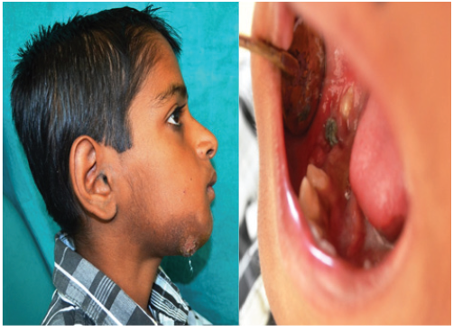
INTRA ORAL IMAGE

ORTHOPANTOMOGRAM IMAGE
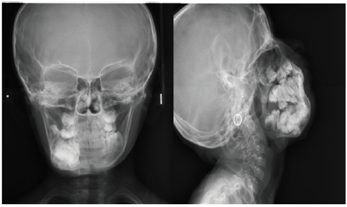
EXTRA ORAL RADIOGRAPHIC IMAGES- PA CEPH & LATERAL OBLIQUE
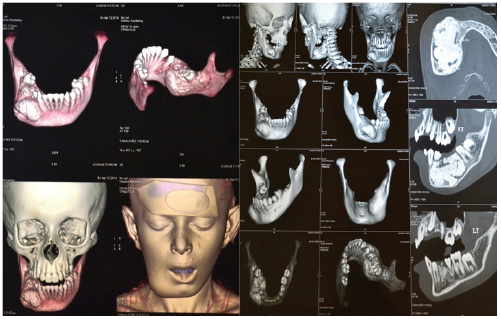
CONTRAT ENHANCED COMPUTED TOMOGRAPHY IMAGES
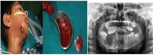
INTRAOPERATIVE & POSTOPERATIVE IMAGE
A 10 year old male patient visited the department of oral medicine and radiology with a chief complaint of pain and swelling on right side of face. Patient gave history of swelling on right side of lower 1/3rd of face since 7-8 years with dull,intermittent pain which aggravates on taking food since 1 month. Patient had undergone radiographic investigations and biopsy on right side of face 15 days back. On extra oral examination, swelling on right side of lower1/3rd of face extending antero-posteriorly from a straight line drawn from inner canthus of eye , posteriorly 1 cm beyond outer canthus of eye. Supero-inferiorly from a straight line drawn from commissure of lip to 1 cm beyond inferior border of mandible. Swelling is approximately 4x4 cm in size. Overlying skin has increase pigmentation with sinus opening, tender on palpation, bony hard. Right submandibular lymph node palpable, mobile and tender. On intra-oral examination, obliteration of vestibule from
44 to 47.Provisional diagnosis of chronic supparative osteomyelitis. OPG reveals dense, homogeneous radio-opaque mass on right side of body with surrounding radiolucent halo extending from 44 to 47. Supero-inferiorly from superior border of mandible to inferior border of mandible to inferior border of mandible with multiple tooth like structures at inferior border of mandible. There is thinning of inferior cortex of mandible with divergence of 43,44,47. CECT reveals a well define lobulated mass with few hypodense areas, measures approximately 37x36x29 mm. the mass is causing significant bicortical expansion of mandible. Radiographic diagnosis- odontomes. Excisional biopsy was performed and histopathologic diagnosis was COMPOUND COMPOSITE ODONTOME.
Disclaimer from Guident
The views and opinions expressed in articles published in Guident are solely those of the authors and do not necessarily reflect the views of the editors, the editorial board or the publisher, Ivory India. Guident does not endorse any product, technique or treatment described. Clinical content is intended for qualified dental and medical professionals and is not a substitute for individual clinical judgement. Guident, its editors and the publisher accept no liability for any consequence arising from the use of information published in the journal.
How to cite this article
Dr. Suraj Agarwal. Clinical Images. Guident. 2017 May;10(6). Available from: https://guident.in/articles/general/clinical-images.html