
Taurodontism
D.Y. Patil Dental College. Mumbai, India
Abstract
Taurodontism can be defined as a change in tooth shape caused by the failure of the Hertwig’s epithelial sheath diaphragm to invaginate at the proper horizontal level. An enlarged pulp chamber, apical displacement of the pulpal floor, and no constriction at the level of the cementoenamel junction are the characteristic features. Endodontic treatment of a taurodont tooth is challenging and requires special handling because of the proximity and apical displacement of the roots. Here, we report a case in which endodontic treatment of the maxillary left first molar with taurodontism was performed.
INTRODUCTION
Taurodontism is a developmental disturbance of a tooth that lacks constriction at the level of the cementoenamel junction (CEJ) and is characterized by vertically elongated pulp chambers, apical displacement of the pulpal floor and bifurcation or trifurcations of the roots 1.
The term “taurodontism” (‘bull tooth’) was coined from the Latin term “tauros”, which means ‘bull’ and the Greek term “odus”, which means ‘tooth’2.
CASE REPORT
A 21-year-old male patient presented at the clinic for endodontic treatment of the maxillary left first molar. Patient had a deep occlusal caries which was tender on percussion. The patient’s medical history was noncontributory. Radiographic examination of the affected tooth revealed an abnormal tooth anatomy.(Fig 1)
The radiographic findings were: The pulp chamber extended beyond the cervical area reaching the furcation. Three short roots were seen at the furcation area in the apical third. From these radiographic findings, the tooth was diagnosed to be a hypertaurodont.
Tooth was isolated under rubber dam & access cavity was prepared. Magnification loupes were used throughout the procedure to facilitate visualization.At the furcation area, three canal orifices were found: palatal, mesiobuccal and distobuccal. A working length radiograph was taken with a #15 file in the canal (Fig 2) and it was confirmed by an electronic apex locator (Root ZX; Morita, Japan). Canals were shaped & cleaned using hand K files & rotary Protaper files. Calcium hydroxide closed dressing was given in 1st appointment & patient was recalled after 4 days.
After drying the canals, AH plus (Dentsply International) sealer was applied onto the root canal walls with a lentulospiral (Dentsply /maillefer; Tulsa, OK, USA). The canals were obturated with lateral condensation. After that, the elongated pulp chamber was obturated with vertical compaction of warm gutta percha. The final radiograph confirmed a well condensed filling of the canals. (Fig 3)

Fig 1: PRE-OPERATIVE VIEW
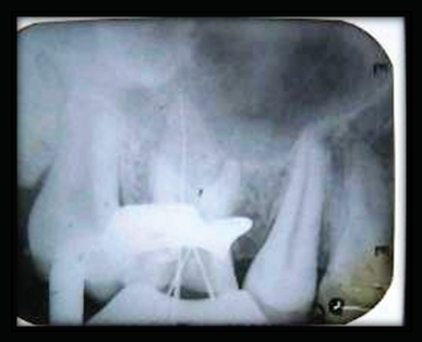
Fig 2: WORKING LENGTH DETERMINATION
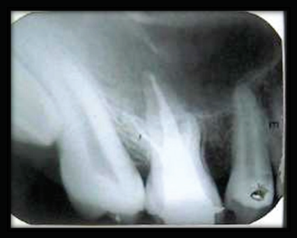
Fig 3: POST-OBTURATION IOPA
DISCUSSION
The etiology of taurodontism is unclear. It is as¬sumed to be caused by the failure of Hertwig’s epithelial diaphragm to invaginate at a suitable horizontal level, resulting in a tooth with short roots, elongated body, an enlarged pulp, and normal dentin.3 Taurodontism has been graded according to its severity: least pronounced (hypotaurodontism), moderate (mesotaurodontism), and most severe (hypertaurodontism).
It was first described by Gorjanovic´-Kramberger (1908); however, the term taurodontism was first coined by Sir Arthur Keith (1913) to describe a molar tooth resembling those of ungulates, particularly bulls.1The prevalence of taurodontism is reported to range from 2.5% to 11.3% of the human population. This range is accounted for by variations in race and differences in diagnostic criteria.11
Taurodontism is associated with several developmental syndromes and anomalies including amelogenesis imperfecta, Down’s syndrome, ectodermal disturbance, Klinefelter syndrome, tricho-dento-osseous syndrome, Mohr syndrome, Wolf-Hirschhorn syndrome and Lowe syndrome8 Its distinguishing features cannot be recognized clinically.4,5 The diagnosis of taurodontism is usually a subjective determination made from diagnostic radio¬graphs. The radiographic characteristics of taurodont tooth are (i) extension of the rectangular pulp chamber into the elongated body of the tooth, (ii) shortened roots and root canals, and (iii) location of furcation near the root apices, despite a normal crown size.4,5
In 1928 Shaw6classified taurodontism arbitrarily, based on relative degree of apical displacement of floor of pulp chamber into hypotaurodontism, meso and hypertaurodontism.
A more effective approach for assessing taurodontism was put forward by Feichtinger and Rossiwall7. They suggested that a tooth can be considered as a taurodont only if the distance from the furcation of the root to the CEJ is greater than the cervico-occlusal distance. Later in 1978 Shiffman and Chanannel8 established mathematical criteria which are adopted by various authors for assessing their cases.
The distance between the baseline connecting the two CEJ and the highest point in the floor of the pulp chamber are used in determining taurodont teeth. Taurodontism is diagnosed in molars when this distance exceeds 2.5 mm.8
A taurodont tooth shows wide variation in the size of the pulp chamber, varying degrees of obliteration and canal configuration, apically positioned canal orifices. Therefore, root canal treatment becomes a challenge 9,10. From an endodontist’s view, taurodontism presents a challenge during negotiation, instrumentation and obturation in root canal therapy. Because of the complexity of the root canal anatomy and proximity of buccal orifices, complete filling of the root canal system in taurodont teeth is challenging. A modified filling technique, which consists of combined lateral compaction in the apical region with vertical compaction of the elongated pulp chamber, has been proposed 9.
REFERENCES
- Jafarzadeh H, Azarpazhooh A, Mayhall JT (2008) Taurodontism: a review of the condition and endodontic treatment challenges. Int Endod J 41, 375-388.
- Keith A (1913) Problems relating to the teeth of the earlier forms of prehistoric man. J R Soc Med 6, 103-124.
- Tsesis I, Shifman A, Kaufman AY. Taurodontism: an endodontic challenge. Report of a case. J Endod. 2003; 29(5): 353-55.
- White SC, Pharoah MJ. Oral Radiology: Principles and Inter¬pretation, 5th Ed. St. Louis: Mosby Inc., 2004: 25.
- Terezhalmy GT, Riley CK, Moore WS. Clinical images in oral medicine and maxillofacial radiology. Taurodontism. Quintes¬sence Int 2001; 32: 254-255.
- Shaw JCM. Taurodont teeth in South African races. J Anat 1928;62: 476-498.
- Feichtinger C, Rosiwall B. Taurodontism in human sex chromosome aneuploidy. Arch Oral Biol 1977; 22:327-329.
- Schiffman A, Chanannel I. Prevalence of taurodontism found in radiographic dental examination of 1200 young adult Israeli patients. Community Dent oral Epidemiol 1978; 6: 200-203.
- Tsesis I, Shifman A, Kaufman AY (2003) Taurodontism: an endodontic challenge: report of a case. J Endod 29, 353-355.
- Rao A, Arathi R (2006) Taurodontism of deciduous and permanent molars: report of two cases. J Indian Soc Pedod Prev Dent 24, 42-44.
More references are availabe on request.
Disclaimer from Guident
The views and opinions expressed in articles published in Guident are solely those of the authors and do not necessarily reflect the views of the editors, the editorial board or the publisher, Ivory India. Guident does not endorse any product, technique or treatment described. Clinical content is intended for qualified dental and medical professionals and is not a substitute for individual clinical judgement. Guident, its editors and the publisher accept no liability for any consequence arising from the use of information published in the journal.
How to cite this article
Dr. Amit Sashte Department of Conservative Dentistry & Endodontics. Taurodontism. Guident. 2015 Aug;8(9). Available from: https://guident.in/articles/endodontics/taurodontism.html


